Презентация Central Nervous System онлайн
На нашем сайте вы можете скачать и просмотреть онлайн доклад-презентацию на тему Central Nervous System абсолютно бесплатно. Урок-презентация на эту тему содержит всего 303 слайда. Все материалы созданы в программе PowerPoint и имеют формат ppt или же pptx. Материалы и темы для презентаций взяты из открытых источников и загружены их авторами, за качество и достоверность информации в них администрация сайта не отвечает, все права принадлежат их создателям. Если вы нашли то, что искали, отблагодарите авторов - поделитесь ссылкой в социальных сетях, а наш сайт добавьте в закладки.
Оцените презентацию от 1 до 5 баллов!
- Тип файла:ppt / pptx (powerpoint)
- Всего слайдов:303 слайда
- Для класса:1,2,3,4,5,6,7,8,9,10,11
- Размер файла:5.70 MB
- Просмотров:101
- Скачиваний:0
- Автор:неизвестен
Слайды и текст к этой презентации:
№1 слайд
Содержание слайда: Central Nervous System
Chapter 13
№2 слайд

Содержание слайда: Introduction
Analogies; telephone switchboard; computer; miracle
A fantastically complex and flexible biological organ
Cephalization become more apparent in higher order species
Increase in the neurons at the rostral end of the CNS
Highest level of cephalization is found in humans
№3 слайд
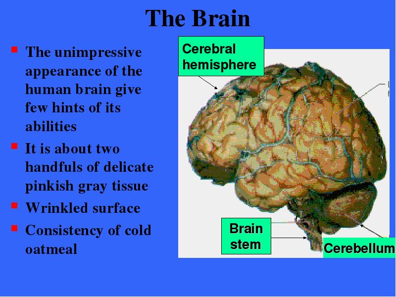
Содержание слайда: The Brain
The unimpressive appearance of the human brain give few hints of its abilities
It is about two handfuls of delicate pinkish gray tissue
Wrinkled surface
Consistency of cold oatmeal
№4 слайд
Содержание слайда: The Brain
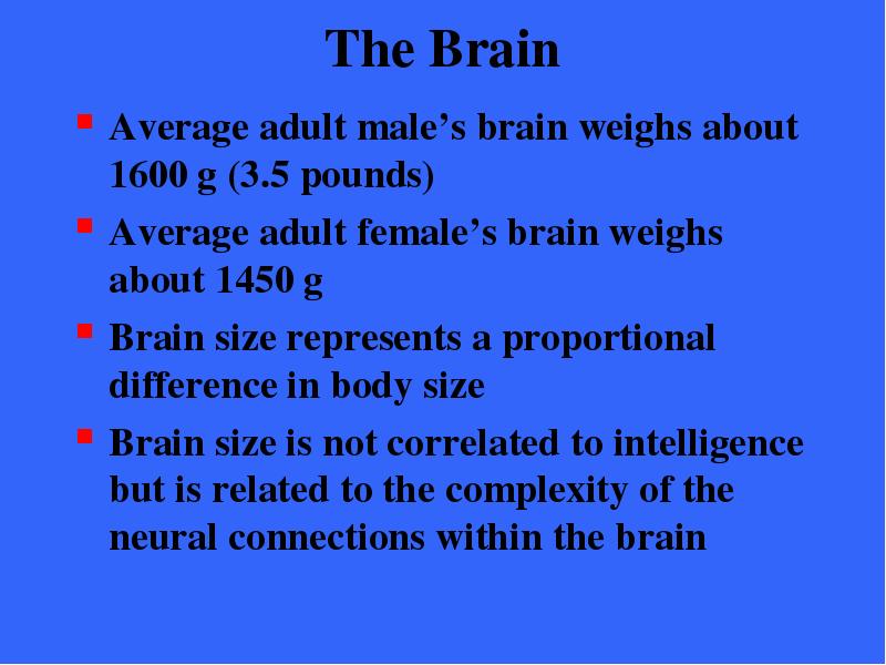
Average adult male’s brain weighs about 1600 g (3.5 pounds)
Average adult female’s brain weighs about 1450 g
Brain size represents a proportional difference in body size
Brain size is not correlated to intelligence but is related to the complexity of the neural connections within the brain
№5 слайд
Содержание слайда: Embryonic Development
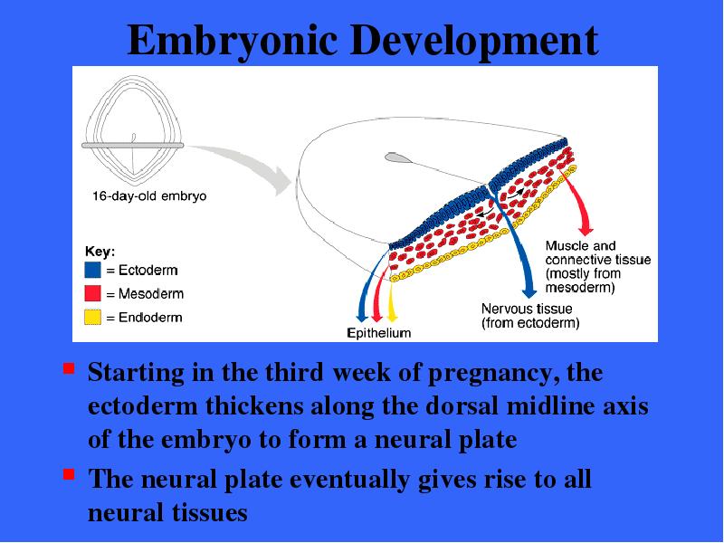
Starting in the third week of pregnancy, the ectoderm thickens along the dorsal midline axis of the embryo to form a neural plate
The neural plate eventually gives rise to all neural tissues
№6 слайд

Содержание слайда: Embryonic Development
The neural plate then invaginates, forming a groove flanked by neural folds
№7 слайд
Содержание слайда: Development of Neural Tube
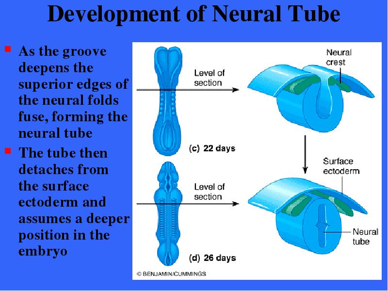
As the groove deepens the superior edges of the neural folds fuse, forming the neural tube
The tube then detaches from the surface ectoderm and assumes a deeper position in the embryo
№8 слайд

Содержание слайда: Development of Neural Tube
The neural tube is formed by the fourth week of pregnancy and differentiates rapidly into the CNS
The brain forms anteriorly and the spinal cord posteriorly
№9 слайд

Содержание слайда: Development of Neural Tube
Small groups of neural fold cells migrate laterally and locate between the surface ectoderm and the neural tube to forming the neural crest
The neural crest gives rise to sensory neurons and some autonomic neurons destined to reside in ganglia
№10 слайд

Содержание слайда: Development of Neural Tube
As soon as the neural tube is formed, its anterior end begins to expand more rapidly than the remaining portion
№11 слайд
Содержание слайда: Primary Brain Vesicles
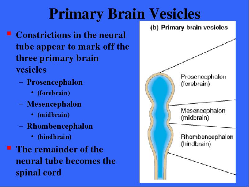
Constrictions in the neural tube appear to mark off the three primary brain vesicles
Prosencephalon
(forebrain)
Mesencephalon
(midbrain)
Rhombencephalon
(hindbrain)
The remainder of the neural tube becomes the spinal cord
№12 слайд
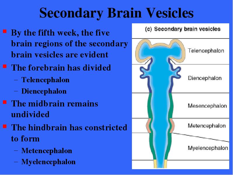
Содержание слайда: Secondary Brain Vesicles
By the fifth week, the five brain regions of the secondary brain vesicles are evident
The forebrain has divided
Telencephalon
Diencephalon
The midbrain remains undivided
The hindbrain has constricted to form
Metencephalon
Myelencephalon
№13 слайд

Содержание слайда: Secondary Brain Vesicles
Each of the five secondary brain vesicles develops rapidly to produce the major structures of the adult brain
The greatest change occurs in the telencephalon which sprouts two large swellings which project anteriorly
These paired expansions become the cerebral hemispheres known collectively as the cerebrum
Hemispheres house ventricles
№14 слайд
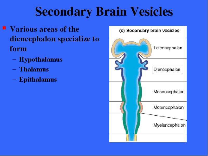
Содержание слайда: Secondary Brain Vesicles
Various areas of the diencephalon specialize to form
Hypothalamus
Thalamus
Epithalamus
№15 слайд

Содержание слайда: Secondary Brain Vesicles
The mesencephalon develops into
Midbrain
Brain stem
№16 слайд

Содержание слайда: Secondary Brain Vesicles
Various areas of the Metencephalon specialize to form
Brain stem
Pons
Cerebellum
№17 слайд

Содержание слайда: Secondary Brain Vesicles
Various areas of the Myelencephalon specialize to form
Brain stem
Medulla oblongata
All the midbrain and hindbrain structures, with the exception of the cerebellum, form portions of the brain stem
№18 слайд
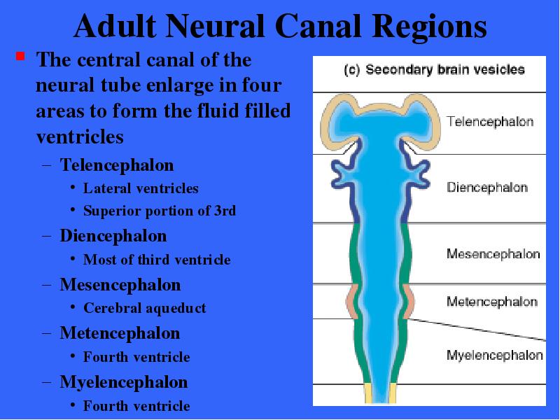
Содержание слайда: Adult Neural Canal Regions
The central canal of the neural tube enlarge in four areas to form the fluid filled ventricles
Telencephalon
Lateral ventricles
Superior portion of 3rd
Diencephalon
Most of third ventricle
Mesencephalon
Cerebral aqueduct
Metencephalon
Fourth ventricle
Myelencephalon
Fourth ventricle
№19 слайд

Содержание слайда: Development of Flexures
During this period of rapid brain growth change is also occurring in the relative position of its parts
Because the brain’s growth is restricted by the skull, midbrain and cervical flexures develop by the fifth week which bend the forebrain toward the brain stem
№20 слайд

Содержание слайда: Effects of Space Restriction
A second consequence of restricted space is that the cerebral hemispheres are forced to take a horseshoe shaped course posteriorly and laterally
Development of the cerebral hemispheres at 13 weeks
№21 слайд

Содержание слайда: Effects of Space Restriction
As a result the hemispheres grow back over and almost completely envelop the diencephalon and midbrain
The cerebral hemispheres at 26 weeks
№22 слайд

Содержание слайда: Effects of Space Restriction
Continued growth of the cerebral hemispheres causes their surfaces to crease and fold
Folding results in convolutions which increase surface area and allow some 1012 neurons to occupy the limited space within the skull
№23 слайд

Содержание слайда: Effects of Space Restriction
The wrinkling of the hemispheres may result from tension on the young axons as they become arranged in a way that minimizes the length of the interconnections they form among the various parts of the cerebrum
№24 слайд

Содержание слайда: Regions of the Brain
The four main regions of the brain are:
Cerebral hemi- spheres
Diencephalon
Thalamus
Hypothalamus
Epithalamus
Brain stem
Midbrain
Pons
Medulla
Cerebellum
№25 слайд

Содержание слайда: Gray and White Matter in CNS
The basic pattern of the CNS can be seen in the spinal cord
A central cavity surrounded by a gray matter core of brain nuclei, external to which is white matter (myelinated fiber tracts)
Figure 12.29 presents major ascending and descending fiber tracts
№26 слайд

Содержание слайда: Gray and White Matter in CNS
The brain has the same basic design except that it also contains additional regions of gray matter that are not evident in the spinal cord
Both the cerebral hemispheres and the cerebellum have an outer layer or cortex of gray matter consisting of neuron cell bodies
№27 слайд

Содержание слайда: Gray and White Matter in CNS
The pattern of white and gray matter changes with descent through the brain stem
The cortex disappears, but scattered gray matter nuclei are seen within the white matter
At the caudal end of the brain stem the basic pattern is evident
№28 слайд

Содержание слайда: Ventricles of the Brain
The ventricles of the brain arise from the expansion of the neural tube
They are continuous with each other and with the central canal of the spinal cord
№29 слайд

Содержание слайда: Ventricles of the Brain
The hollow ventricular chambers are filled with cerebrospinal fluid and lined by ependymal cells
№30 слайд

Содержание слайда: Ventricles of the Brain
The paired lateral ventricles are large C-shaped chambers that reflect the pattern of cerebral growth
One lateral ventricle is located in each cerebral hemisphere
№31 слайд

Содержание слайда: Ventricles of the Brain
Anteriorly, the lateral ventricles lie close together separated only by a thin median membrane called the septum pellucidum
Each ventricle communicates with the narrow third ventricle in the diencephalon
№32 слайд

Содержание слайда: Ventricles of the Brain
Communication occurs through the inter- ventricular foramen (foramen of Moro)
№33 слайд

Содержание слайда: Ventricles of the Brain
The third ventricle is continuous with the fourth ventricle via the canal-like cerebral aqueduct that runs through the midbrain
№34 слайд
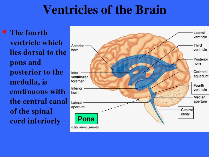
Содержание слайда: Ventricles of the Brain
The fourth ventricle which lies dorsal to the pons and posterior to the medulla, is continuous with the central canal of the spinal cord inferiorly
№35 слайд

Содержание слайда: Ventricles of the Brain
Three openings mark the walls of the fourth ventricle
Paired lateral apertures
Median aperture
Apertures connect the ventricles to the subarachnoid space
№36 слайд

Содержание слайда: The Cerebral Hemispheres
The cerebral hemispheres form the superior part of the brain
These two structures account for about 83% of the mass of the brain
The two hemispheres cover and obscure the diencephalon and the top of the brain stem
№37 слайд

Содержание слайда: The Cerebral Hemispheres
Nearly the entire surface of the cerebral hemispheres is marked by elevated ridges of tissues called gyri separated by shallow grooves called sulci
Deeper grooves called fissures separate larger regions of the brain
№38 слайд

Содержание слайда: The Cerebral Hemispheres
Prominent gyri and sulci are similar in all people
The median longitudinal fissure separates the hemispheres
The transverse fissure separates the cerebral hemispheres from the cerebellum below
№39 слайд

Содержание слайда: Lobes of Cerebral Hemispheres
Deeper sulci divide each hemisphere into five lobes
Frontal lobe
Temporal lobe
Parietal lobe
Occipital lobe
Insula (located within the lateral sulcus)
№40 слайд

Содержание слайда: Lobes of Cerebral Hemispheres
Location of the insula deep within the Lateral sulcus of the hemisphere
№41 слайд

Содержание слайда: Fissures of Cerebral Hemispheres
Sulci divide lobes of the hemispheres
Central sulcus
Parieto- occipital sulcus
Lateral sulcus
Transverse fissure
№42 слайд

Содержание слайда: Medial Surface of Right Hemisphere
Medial surface of the right hemisphere showing the Parieto- occipital sulcus
№43 слайд

Содержание слайда: Position of Cerebral Hemispheres
The frontal lobes occupy the anterior cranial fossa
The anterior parts of the temporal lobes fill the middle cranial fossa
The cerebellum and brain stem occupies the posterior cranial fossa and the occipital lobes rests upon it
№44 слайд

Содержание слайда: Cerebral Cortex
The cerebral cortex is the “executive suite” of the nervous system
It enables us to perceive, communicate, remember, understand, appreciate, and initiate voluntary movements
Literally all qualities associated with conscious behavior or consciousness originate within the cerebral cortex of the various lobes of the brain
№45 слайд

Содержание слайда: Cerebral Cortex
The cerebral cortex is gray matter composed of neuron cells bodies, dendrites, and unmyelinated axons (plus support cells and blood vessels)
It is only 2-4 mm thick
The many convolutions of the brain effectively triple its surface area
It accounts for roughly 40% of total brain mass
№46 слайд
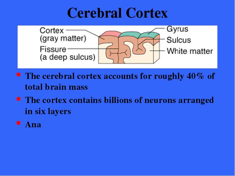
Содержание слайда: Cerebral Cortex
The cerebral cortex accounts for roughly 40% of total brain mass
The cortex contains billions of neurons arranged in six layers
Ana
№47 слайд

Содержание слайда: Cerebral Hemispheres
Coronal section of the brain which reveals the cerebral cortex, white matter, and basal nuclei within the cerebral hemispheres
№48 слайд

Содержание слайда: Cerebral Cortex
Research on the structure and function of the brain reveals that there are both specialized and diffuse areas of function
Motor and sensory areas are localized in discrete cortical areas called domains
Many higher mental functions such as memory and language appear to have overlapping domains and are more diffusely located
Broadmann areas are areas of localized function
№49 слайд

Содержание слайда: Cerebral Cortex - Generalizations
The cerebral cortex has three types of functional areas
Motor areas / control voluntary motor function
Sensory areas / provide conscious awareness of sensation
Association areas / act mainly to integrate diverse information for purposeful action
Each hemisphere is chiefly concerned with the sensory and motor functions of the opposite (contralateral) side of the body
№50 слайд

Содержание слайда: Cerebral Cortex - Generalizations
Although they are largely symmetrical in structure the two hemispheres are not entirely equal in function, instead there is lateralization of cortical function
Remember that the information presented is a gross oversimplification to convey and clarify concepts
№51 слайд

Содержание слайда: Motor Areas
Cortical areas controlling motor functions lie in the posterior part of the frontal lobes
Motor areas include the primary motor cortex, the premotor cortex, Broca’s area, and the front eye field
№52 слайд

Содержание слайда: Primary Motor Cortex
The primary motor cortex is located in the precentral gyrus of the frontal lobe of each hemisphere
Large neurons (pyramidal cells) in these gyri allow us to consciously control the precise or skill voluntary movements of our skeletal muscles
№53 слайд

Содержание слайда: Pyramidal cells
These long axons, which project to the spinal cord, form the massive voluntary motor tracts called the pyramidal, or corticospinal tracts
All other descending motor tracts issue from brain stem nuclei and consists of chains of two, three, or more neurons
№54 слайд

Содержание слайда: Pyramidal Tracts
The lateral corticospinal tract consists of the long axons of the pyramidal cells located within the primary motor cortex
№55 слайд

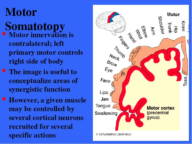
Содержание слайда: Motor Somatotopy
Body is represented spatially in the primary motor cortex of each hemisphere
Most of the neurons in these gyri control muscles in body areas having the most precise motor control
The areas with the most control (face, tongue, and hands)
№56 слайд
Содержание слайда: Motor Somatotopy
Motor innervation is contralateral; left primary motor controls right side of body
The image is useful to conceptualize areas of synergistic function
However, a given muscle may be controlled by several cortical neurons recruited for several specific actions
№57 слайд

Содержание слайда: Motor Somatotopy
Damage to the localized areas of the primary motor cortex paralyzes the muscles controlled by this area
If the lesion is in the right hemisphere, the left side will be paralyzed
Only voluntary control is lost as the muscles can still contract reflexively
№58 слайд

Содержание слайда: Premotor Cortex
The premotor cortex controls motor skills of repetitive or patterned nature (typing or piano)
The premotor cortex coordinates the movement of several muscle groups to act simultaneously or sequentially
№59 слайд

Содержание слайда: Premotor Cortex
The premotor cortex sends activating impulses to the primary motor cortex
Also influences motor actively more directly by supplying about 15% of pyramidal tract fibers
A memory bank of skilled motor activities
№60 слайд

Содержание слайда: Premotor Cortex
This area appears to involved with motor planning
It controls voluntary actions that depend on sensory feedback
№61 слайд

Содержание слайда: Premotor Cortex
Damage to the premotor area results in the loss of the motor skills in that region
Muscle strength and the ability to perform the discrete individual movements are not hindered
Neurons relearning the skill would require practice
№62 слайд

Содержание слайда: Broca’s area
The area has long been considered to be present in only one hemisphere (usually left)
A special motor speech area that directs the muscles of the tongue, throat, and lips in articulating words
№63 слайд

Содержание слайда: Broca’s area
Recent PET scans indicates that Broca’s area and a similar area in the opposite hemisphere become active as we prepare to speak
The areas may be involved with planning speech and other voluntary motor activities
№64 слайд
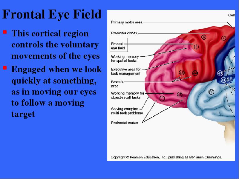
Содержание слайда: Frontal Eye Field
This cortical region controls the voluntary movements of the eyes
Engaged when we look quickly at something, as in moving our eyes to follow a moving target
№65 слайд

Содержание слайда: Sensory Areas
Areas concerned with the conscious awareness of sensation in the parietal, temporal and occipital lobes
№66 слайд
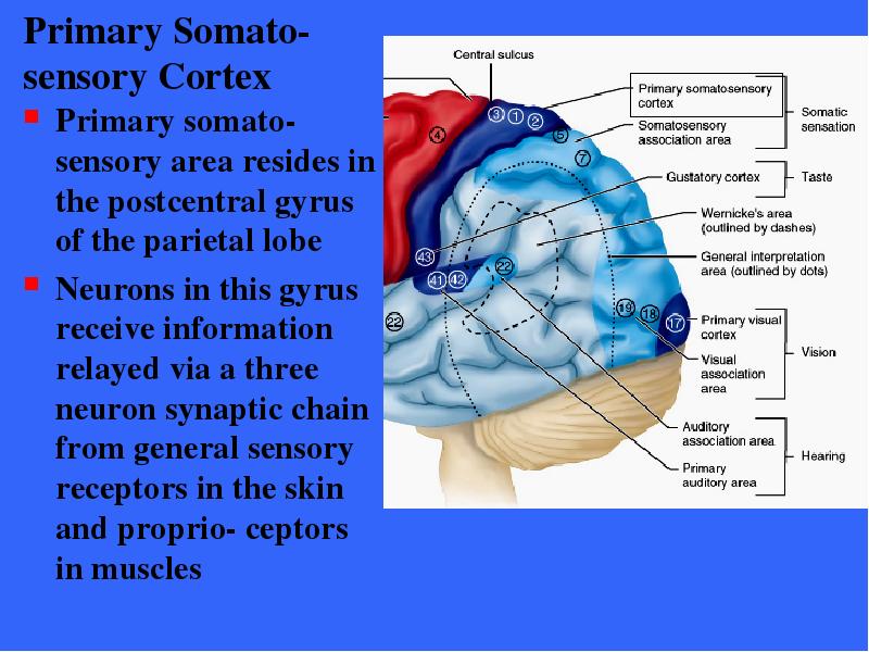
Содержание слайда: Primary Somato-sensory Cortex
Primary somato- sensory area resides in the postcentral gyrus of the parietal lobe
Neurons in this gyrus receive information relayed via a three neuron synaptic chain from general sensory receptors in the skin and proprio- ceptors in muscles
№67 слайд

Содержание слайда: Synaptic Chain
Central axons of sensory (1st order) neurons enter dorsal root of spinal cord
Synapse with 2nd order neurons in medial lemniscal tract and ascend to Thalamus
Synapse with 3rd order neurons which transmit to somato- sensory cortex
№68 слайд

Содержание слайда: Primary Somato-sensory Cortex
In the cortex neurons process the sensory information and identify the precise area of the body being stimulated
This ability to localize (assign a location) a stimulus precisely is called spatial discrimination
№69 слайд
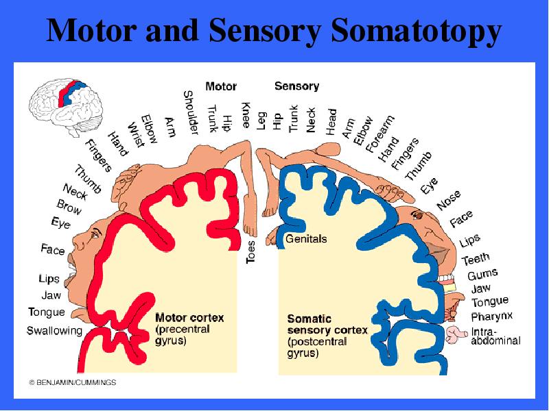
Содержание слайда: Motor and Sensory Somatotopy
№70 слайд

Содержание слайда: Primary Somato-sensory Cortex
The sensory spatial discrimination is contralateral with the right hemisphere receiving inputs from the left side of the body
The entire body is represented spatially in the primary somatosensory area of each hemisphere
№71 слайд

Содержание слайда: Primary Somato-sensory Cortex
The amount of sensory cortex devoted to a particular body region is related to how many sensory receptors are present
In humans the face (especially the lips) and fingertips are the most sensitive body areas
№72 слайд

Содержание слайда: Primary Somatosensory Cortex
Damage to the primary somatisensory cortex destroys the conscious ability to feel and localize touch, pressure, and vibrations on the skin
Most ability to feel pain and temperature is also lost, although these can still be felt in a vague, poorly localized way
№73 слайд

Содержание слайда: Somatosensory Association Area
The area lies just posterior to the primary somata- sensory cortex and has many connections with it (Broadmann 5,7)
The major function of the area is to integrate and analyze different somatic sensory inputs (touch, pressure, others) relayed to it by the primary somato- sensory cortex
№74 слайд

Содержание слайда: Somatosensory Association Area
The somatosensory association area forms a comprehensive evaluation of what is being felt relative to its size, texture and parts
The somatosensory association area draws upon stored memories of past sensory experiences to perceive the object as one you recognize
№75 слайд

Содержание слайда: Somatosensory Association Area
Past associations allow you to recognize familiar objects (coins, keys) without having to look at them
Someone with damage to this area would not be able to recognize what they are feeling without actually looking at the object
№76 слайд

Содержание слайда: Primary Visual Cortex
The primary visual cortex (17) is located on the posterior and medial portions of the occipital lobe
№77 слайд

Содержание слайда: Primary Visual Cortex
Most of the primary visual cortex is located on the medial aspect of the occipital lobe buried within the deep calcarine sulcus
№78 слайд

Содержание слайда: Primary Visual Cortex
The largest of all cortical sensory areas, the primary visual cortex receives visual information that originates on the retinas of the eyes
There is a map of visual space on the primary visual cortex analogous to the body map of the somato- sensory cortex
№79 слайд

Содержание слайда: Primary Visual Cortex
Again, the right half of visual space is represented on the left visual cortex, the left half on the right cortex
If this cortical area is damaged, the person has no conscious awareness of what is being viewed and is functionally blind
№80 слайд

Содержание слайда: Primary Visual Cortex
The primary visual cortex is the first of a series of cortical areas that process visual input
The processing here is at a comparatively low level - noting the orientation of objects being viewed and putting the inputs from the two eyes together
№81 слайд

Содержание слайда: Visual Association Area
This area surrounds the primary visual area and encompasses much of the occipital lobe (18, 19)
Communicating with the primary visual area, the visual association area continues the processing of visual information
№82 слайд

Содержание слайда: Visual Association Area
This area analyzes color, form and movement in light of past visual experiences so that we might recognize & appreciate what we are seeing
№83 слайд
Содержание слайда: Visual Association Area
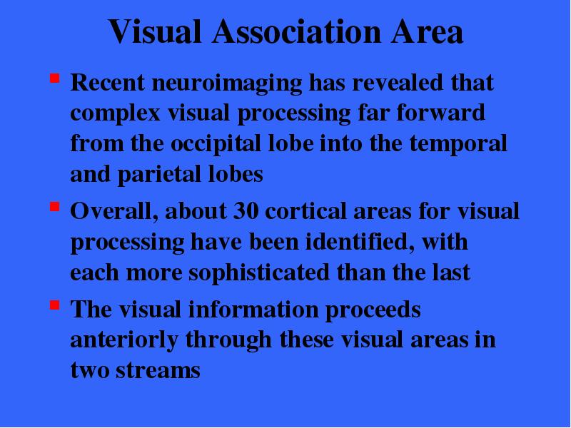
Recent neuroimaging has revealed that complex visual processing far forward from the occipital lobe into the temporal and parietal lobes
Overall, about 30 cortical areas for visual processing have been identified, with each more sophisticated than the last
The visual information proceeds anteriorly through these visual areas in two streams
№84 слайд

Содержание слайда: Visual Association Area
The ventral stream extends through the inferior part of the entire temporal lobe and is responsible for recognizing objects, words during reading, and faces
Facial recognition is right hemisphere only
The “what”
№85 слайд

Содержание слайда: Visual Association Area
The dorsal stream extends through the posterior parietal cortex to the postcentral gyrus and perceives spatial relationships among different objects
The “where” things are in space
№86 слайд

Содержание слайда: Visual Association Area
The dorsal stream in the parietal lobe is important for spatial perception
The superior part of the lobe calculates how we move our limbs through space then sends this information to the motor cortex which dictates these movements
In addition, the parietal lobe is important for abstract mathematical abilities, which are highly visual, spatial in nature
№87 слайд

Содержание слайда: Visual Areas
Damage to the visual cortex results in functional blindness
Damage to the visual association areas results in an ability to see but not comprehend what is seen
№88 слайд

Содержание слайда: Primary Auditory Cortex
The primary auditory cortex is located on the superior margin of the temporal lobe, primarily inside the lateral sulcus
Broadmann 41,42
It provides us with our conscious awareness of sound
№89 слайд

Содержание слайда: Primary Auditory Cortex
Hearing receptors in the cochlear of the inner ear transmit impulses to primary auditory cortex
Impulses related to loudness, rhythm, and especially pitch (high to low notes) is complied
№90 слайд

Содержание слайда: Auditory Association Area
The auditory association area lies just posterior to the primary auditory area Broadmann 22
This area evaluates and classifies sound
Memories of past sounds seem to be stored here
№91 слайд

Содержание слайда: Auditory Association Area
In one hemisphere (usually the left), the auditory association areas lies in the center of Wernicke’s area
This functional area is involved in recognizing and understanding spoken words
№92 слайд

Содержание слайда: Auditory Association Area
Damage to Wernicke’s area interferes with the ability to comprehend speech
№93 слайд
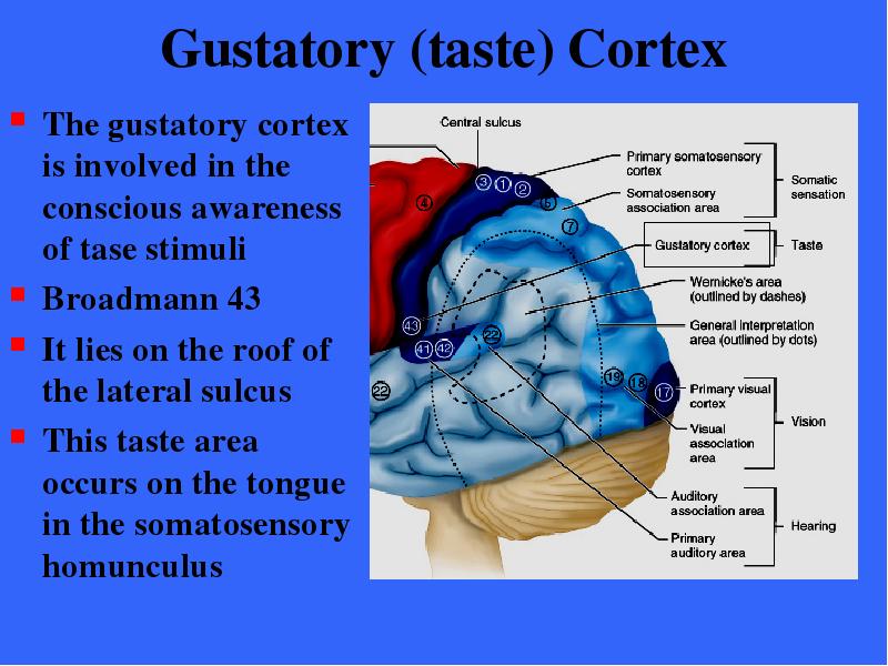
Содержание слайда: Gustatory (taste) Cortex
The gustatory cortex is involved in the conscious awareness of tase stimuli
Broadmann 43
It lies on the roof of the lateral sulcus
This taste area occurs on the tongue in the somatosensory homunculus
№94 слайд

Содержание слайда: Vestibular (equilibrium) Cortex
The cortex is responsible for conscious aware-ness of the sense of balance; specifically the position of the head in space
Recent studies have placed this region in the posterior insula deep in the lateral sulcus
№95 слайд

Содержание слайда: Olfactory Area
The primary olfactory cortex lie on the medial aspects of the cerebrum in a small region called the piriform lobe of which the hook-like uncus is the dominant feature
№96 слайд

Содержание слайда: Olfactory Area
The olfactory nerves (Cranial nerve I) from the nasal cavity transmit impulses that are ultimately relayed to the olfactory cortex
The outcome is conscious awareness of smells
№97 слайд

Содержание слайда: Olfactory Area
The olfactory cortex is part of a brain area called the rhinencephalon (nose brain) which includes all parts of the cerebrum that directly receive olfactory signals
№98 слайд

Содержание слайда: Olfactory Area
The piriform lobe, the olfactory tract, the olfactory bulb, and some nearby structures are all components of the rhinencephalon
№99 слайд

Содержание слайда: Olfactory Area
The rhinencephalon connects to the brain area that is involved in emotions, the limbic system, which explains why smells often trigger emotions
№100 слайд

Содержание слайда: Olfactory Area
Part of the frontal lobe, the orbitofrontal cortex, is involved in higher-order processing of smells
Consciously identifying and recalling specific odors and telling different smells apart
№101 слайд
Содержание слайда: Association Areas
Association areas include all cortical areas other than primary sensory and motor areas
The name reflects the fact that some of these areas tie together, or make associations between different kinds of sensory information
They also seem to associate new sensory inputs with memories of past experiences
№102 слайд
Содержание слайда: Association Areas
The term association area is fading from use and will probably be replaced by higher- order processing areas
Higher-order processing areas is a more accurate name as these areas, which are nearby the primary sensory areas, have the ability to analyze, recognize, and act on the sensory input received
№103 слайд

Содержание слайда: Prefrontal Cortex
The prefrontal cortex occupies the large region of the frontal lobe anterior to the motor area
The most complicated cortical region
It performs many cognitive functions
№104 слайд

Содержание слайда: Prefrontal Cortex
Cognition is all aspects of thinking, perceiving and of intentionally remembering and recalling information
The prefrontal cortex is necessary for abstract ideas, reasoning and judgment, impulse control, persistence, long term planning
№105 слайд

Содержание слайда: Prefrontal Cortex
The prefrontal cortex also is used for long- term planning, complex problem solving, mental flexibility, social skills, appreciating humor, empathy, and conscience
№106 слайд

Содержание слайда: Prefrontal Cortex
The prefrontal cortex also seems to be related to mood and has close links to the emotional (limbic) part of the forebrain
Tumors in this region may cause mental and personality disorders
The tremendous elaboration of this prefrontal region distinguishes humans from animals
№107 слайд
Содержание слайда: Prefrontal Cortex
Functional neuro-imaging techniques have begun to reveal the functions of specific parts of the prefrontal cortex
Completion of multi-step problem solving tasks requires the temporary storage of information in working memory
№108 слайд

Содержание слайда: Prefrontal Cortex
The working memories of spatial relations are stored in the dorsolateral prefrontal cortex just anterior to the frontal eye field
№109 слайд

Содержание слайда: Prefrontal Cortex
Working memories of objects and faces are stored farther ventrally, below Broca’s area
№110 слайд

Содержание слайда: Prefrontal Cortex
More significant is the region that manages cognitive tasks by directing our attention to the relevant information in the working memory
This executive area lies between the working- memory sites, just anterior to Broca’s area
№111 слайд

Содержание слайда: Prefrontal Cortex
The extreme anterior pole of the frontal cortex was found to be active in solving the most complex problems - problems in which many sub- problems had to be completed before a solution could be obtained
№112 слайд

Содержание слайда: Prefrontal Cortex
The new findings suggest support for a general rule of neuroscience that says the farther rostrally one goes in the CNS, the more complex are the neuron functions performed
№113 слайд
Содержание слайда: Prefrontal Cortex
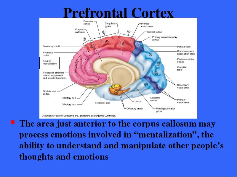
The area just anterior to the corpus callosum may process emotions involved in “mentalization”, the ability to understand and manipulate other people’s thoughts and emotions
№114 слайд

Содержание слайда: General Interpretation Area
The existence of this area within the brain is debated
Once thought to be an area of integration of all types of sensory information, its existence was mainly substantiated by agnosia (not knowing)
Recent studies do not support its presence
№115 слайд

Содержание слайда: Language Area
The large area surrounding the lateral sulcus in the left cerebral hemisphere is involved in various functions related to language
№116 слайд

Содержание слайда: Language Area
Five areas have been identified with language; Broca’s area (speech production); Wernicke’s area (speech comprehension); prefrontal cortex (conceptual analysis); temporal lobe (visual and auditory aspects of language ); the insula (recognition of rhythms)
№117 слайд

Содержание слайда: Language Area
The corresponding areas on the right hemisphere, although not involved in the mechanics of language, act in the creative interpretation of words and in controlling the emotional overtones of speech
№118 слайд

Содержание слайда: Insula
The insula is large and the functions of its cortex are not well understood
Some parts function in language and some in the sense of balance
Other parts have visceral function including the perception of upset stomach, full bladder
№119 слайд

Содержание слайда: Lateralization of Cortical Function
We use both cerebral hemispheres for almost every task and it appears the hemispheres share memories and appear nearly identical
However, there are differences and unique abilities that are found in one hemisphere and not the other
This phenomenon is call lateralization
Cerebral dominance suggest that there is one hemisphere that dominates each task
№120 слайд

Содержание слайда: Lateralization of Cortical Function
In most people (Approx. 90%) the left hemisphere has greater control over language abilities, mathematical abilities, and logic
The other hemisphere (usually the right) is involved in visual-spatial skills, intuition, emotion, and appreciation of art and music
№121 слайд
Содержание слайда: Lateralization of Cortical Function
Most individuals (90%) with left cerebral dominance are right-handed
In the remaining 10% the roles of the hemispheres are reversed or the hemispheres share their functions equally
Typically, many right cerebral dominant people are left handed and more often male
In lefties the cerebral cortex functions bilaterally, the mutuality of brain control sometimes result in ambidexterity or dyslexia
№122 слайд

Содержание слайда: Lateralization of Cortical Function
The two cerebral hemispheres have perfect and almost instantaneous communication with one another via connecting fiber tracts as well as complete integration of their functions
Lateralization means that each hemisphere is better than the other at certain functions, neither side is better at everything
№123 слайд

Содержание слайда: Cerebral White Matter
Communication within the brain is extensive
The cerebral white matter deep to the gray matter of the cortex provides for communication between cerebral areas and between the cortex and the lower CNS centers
№124 слайд

Содержание слайда: Cerebral White Matter
The white matter largely consists of myelinated fibers bundled into large tracts
These fibers and the tracts they form are classified according to the direction in which they run as
Commissural
Association
Projection
№125 слайд

Содержание слайда: Cerebral White Matter
Commissures connect the hemispheres
Association fibers connect areas within hemispheres
Projection tracts connect higher & lower areas of CNS
№126 слайд

Содержание слайда: Cerebral White Matter
Commissures connect the corresponding areas of two hemispheres enabling them to function as a whole
The Corpus callosum is the largest commissure
№127 слайд
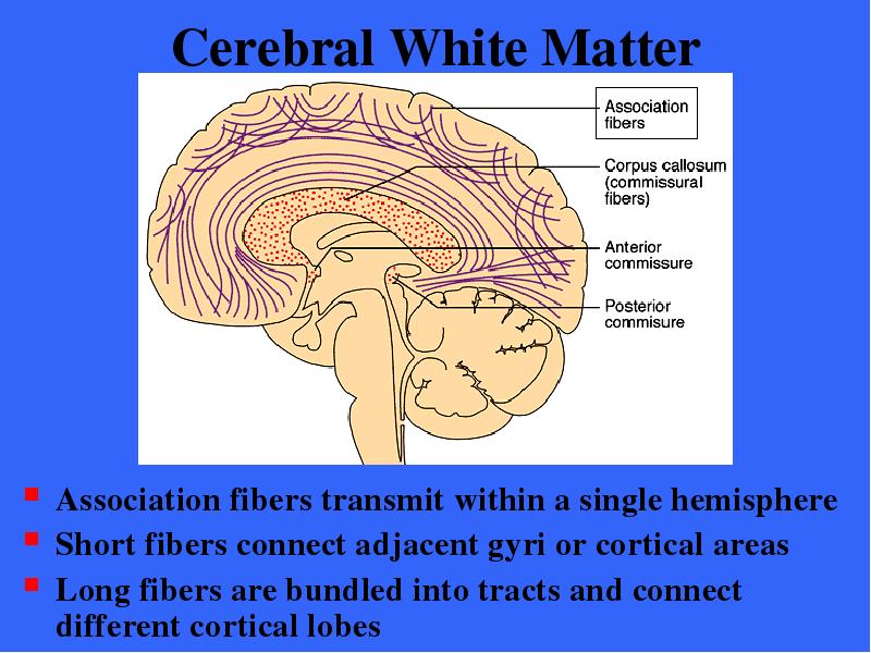
Содержание слайда: Cerebral White Matter
Association fibers transmit within a single hemisphere
Short fibers connect adjacent gyri or cortical areas
Long fibers are bundled into tracts and connect different cortical lobes
№128 слайд
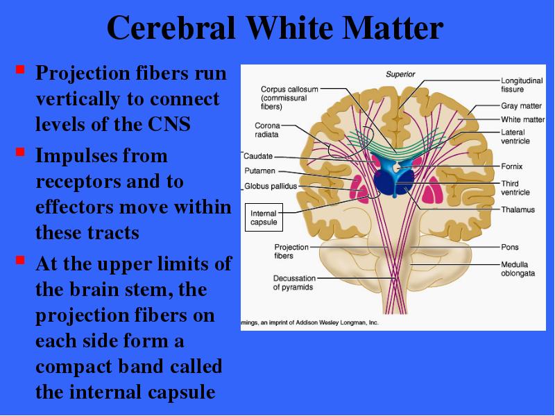
Содержание слайда: Cerebral White Matter
Projection fibers run vertically to connect levels of the CNS
Impulses from receptors and to effectors move within these tracts
At the upper limits of the brain stem, the projection fibers on each side form a compact band called the internal capsule
№129 слайд

Содержание слайда: Cerebral White Matter
Ascending projection tracts pass between the thalamus and the basal nuclei beyond which the radiate through the cerebral white matter to the cortex
This distinctive arrangement of projection tract fibers is called the corona radiata
№130 слайд

Содержание слайда: Cerebral White Matter
The fibers of the corona radiata fan out into the white matter of the cerebral hemisphere
№131 слайд

Содержание слайда: Basal Nuclei
In the cerebral white matter of each hemisphere are a groups of subcortical nuclei called the basal nuclei
The main mass of this tissue include the caudate nucleus, putamen, and the globus pallidus
№132 слайд

Содержание слайда: Basal Nuclei
The putamen and globus pallidus together form a mass called the lentiform nucleus
№133 слайд
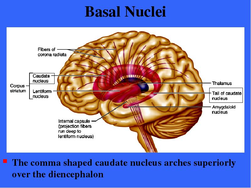
Содержание слайда: Basal Nuclei
The comma shaped caudate nucleus arches superiorly over the diencephalon
№134 слайд

Содержание слайда: Basal Nuclei
The lentiform nucleus flanks the internal capsule laterally
№135 слайд
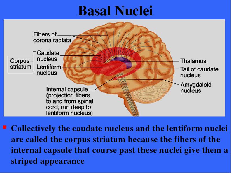
Содержание слайда: Basal Nuclei
Collectively the caudate nucleus and the lentiform nuclei are called the corpus striatum because the fibers of the internal capsule that course past these nuclei give them a striped appearance
№136 слайд

Содержание слайда: Basal Nuclei
The basal nuclei are functionally associated with the subthalamic nuclei (located in the floor of the diencephalon) and the substantia nigra of the midbrain
№137 слайд

Содержание слайда: Basal Nuclei
The amygdaloid nucleus sits on the tail of the caudate nucleus, functionally it belongs to the limbic system
№138 слайд

Содержание слайда: Basal Nuclei
Functionally, the basal nuclei can be viewed as complex neural calculators that cooperate with the cerebral cortex in controlling movement
№139 слайд

Содержание слайда: Basal Nuclei
The basal nuclei receive inputs from the entire cerebral cortex as well as from other subcortical nuclei
Via relays through the thalamus, the basal nuclei project to the premotor and prefrontal cortices
№140 слайд

Содержание слайда: Basal Nuclei
Via relays the basal nuclei influence muscle movements directed by the primary motor cortex
The basal nuclei has no direct access to the motor pathways
The precise role of the basal nuclei is difficult to determine since their function overlaps to some extent with the cerebellum
The basal nuclei are particularly important in starting, stopping, and monitoring movements executed by the by the cortex
№141 слайд

Содержание слайда: Basal Nuclei
The nuclei are involved in monitoring muscle movements that are relatively slow and sustained or patterned
The nuclei also regulated the intensity of these movements
Additionally, they inhibit antagonistic or unnecessary movements
When the basal nuclei are impaired, the result is disturbances in posture and muscle tone, involuntary movements including tremors, and abnormal slowness
№142 слайд

Содержание слайда: The Diencephanlon
Forms the central core of the forebrain and is surrounded by the cerebral hemispheres
№143 слайд

Содержание слайда: The Diencephalon
The diencephalon consists of three structures
Thalamus
Hypothalamus
Epithalamus
These structures effectively enclose the third ventricle
№144 слайд

Содержание слайда: The Diencephalon
The three structures of the diencephalon
Thalamus
Hypothalamus
Epithalamus
These structures are shown with the hemispheres removed
№145 слайд
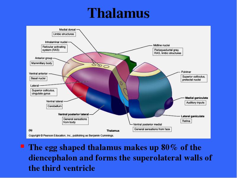
Содержание слайда: Thalamus
The egg shaped thalamus makes up 80% of the diencephalon and forms the superolateral walls of the third ventricle
№146 слайд

Содержание слайда: Thalamus
The thalamus is composed of bilateral masses of gray matter held together by a mid- line commissure called the intermediate mass
№147 слайд

Содержание слайда: Thalamus
The thalamus has many different nuclei, most named for their location
Each of these nuclei has a functional specialization
Each projects fibers to and receives fibers from a specific region of the cerebral cortex
№148 слайд

Содержание слайда: The Thalamus
Sensory inputs are not the only type of information relayed through the thalamus
Every part of the brain that communicates with the cerebral cortex must relay signals through the nucleus of the thalamus
The thalamus can therefore be thought of as the gateway to the cerebral cortex
№149 слайд

Содержание слайда: Thalamus
Afferent impulses from all senses and all parts of the body converge on the thalamus and synapse with at least one of its nuclei
Within the thalamus, a sorting-out and information “editing” process occurs
№150 слайд

Содержание слайда: Thalamus
Impulses having to do with similar functions are grouped together and relayed via the internal capsule to the appropriate area of the sensory cortex as well as specific cortical association areas
№151 слайд

Содержание слайда: Thalamus
In addition to sensory inputs, virtually all inputs ascending to the cerebral cortex funnel through thalamic nuclei
Ventral posterior lateral nucleus
General somatic sensory receptors (touch, pain pressure)
№152 слайд
Содержание слайда: Thalamus
Lateral geniculate body
Visual relay from retina
Medial geniculate body
Auditory inputs
Anterior nuclear group
Regulation of emotion and visceral function
Ventral lateral nuclei
Direct motor activity of cerebellum
Ventral anterior nuclei
Direct motor activity of basal nuclei
№153 слайд

Содержание слайда: Thalamus
Pulvinar, medial dorsal and lateral nuclei are involved in the integration of sensory information and projection to specific association cortices
№154 слайд

Содержание слайда: Thalamus
The thalamus plays a key role in mediating sensation, motor activities, cortical arousal, learning, and memory
It is truly the gateway to the cerebral cortex
№155 слайд

Содержание слайда: The Hypothalamus
The hypothalamus is located below the thalamus, capping the brain stem
№156 слайд

Содержание слайда: Hypothalamus
Merging into the midbrain inferiorly, it extends from the optic chiasma to the posterior margin of the mammillary bodies
№157 слайд
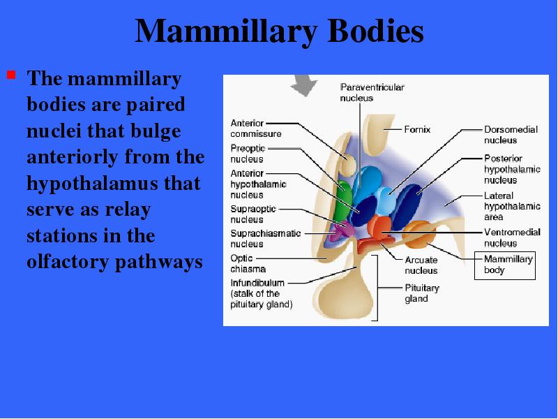
Содержание слайда: Mammillary Bodies
The mammillary bodies are paired nuclei that bulge anteriorly from the hypothalamus that serve as relay stations in the olfactory pathways
№158 слайд

Содержание слайда: Hypothalamus
Between the optic chiasma and the mammillary bodies is the infundibulum
A stalk of the hypothalamic tissue connects the pituitary gland to the base of hypothalamus
№159 слайд

Содержание слайда: Hypothalamus
The hypothalamus contains about a dozen functionally important nuclei
Despite its small size, the hypothalamus is the main visceral control center of the body and is vitally important to overall body homeostasis
№160 слайд

Содержание слайда: Autonomic Control Center
The hypothalamus regulates involuntary nervous activity by controlling the activity of autonomic centers in the brain stem and spinal cord
In this role the hypothalamus influences
Blood pressure
Rate and force of heart contraction
Motility of the digestive system
Respiratory rate and depth
Secretion of sweat and salivary glands
№161 слайд

Содержание слайда: Center for Emotional Response
The hypothalamus has numerous connections with cortical association areas, lower brain stem centers, and it lies at the center of the limbic system which is the emotional part of the brain
Nuclei involved in the perception of fear, pleasure, and rage, as well as those involved in the biological rhythms and drives of sex are found in the hypothalamus
№162 слайд
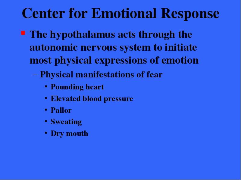
Содержание слайда: Center for Emotional Response
The hypothalamus acts through the autonomic nervous system to initiate most physical expressions of emotion
Physical manifestations of fear
Pounding heart
Elevated blood pressure
Pallor
Sweating
Dry mouth
№163 слайд

Содержание слайда: Body Temperature Regulation
The body’s thermostat is in the hypothalamus
The hypothalamus receives input from the thermoreceptors located in other parts of the brain as well as in the body periphery
Homeostatic adjustments are then made to either cool or heat the body (sweating or shivering)
Hypothalamic centers also induce fever
№164 слайд

Содержание слайда: Body Temperature Regulation
Hypothalamic receptors in the preoptic region monitor the temperature of the blood flowing through the hypothalamus
№165 слайд
Содержание слайда: Body Temperature Regulation
According to signals received by the preoptic nuclei the hypothalamus initiates mechanisms to maintain relatively constant body temperature
Cooling / sweating
Heat generation / shivering
№166 слайд
Содержание слайда: Regulation of Hunger & Thirst
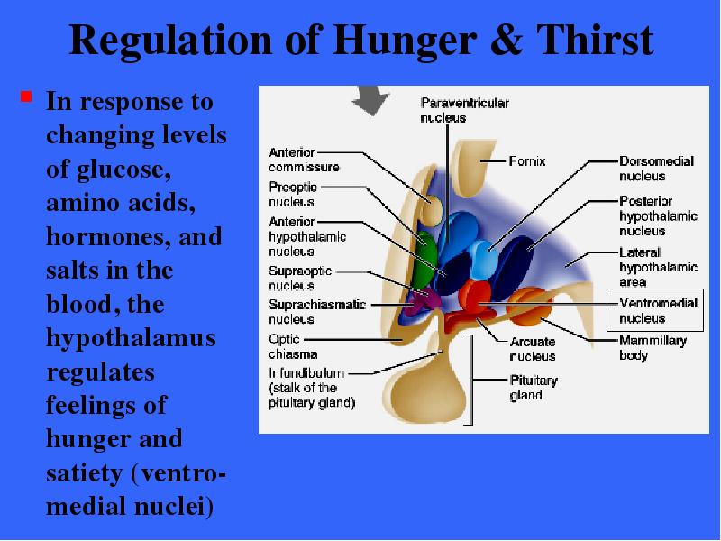
In response to changing levels of glucose, amino acids, hormones, and salts in the blood, the hypothalamus regulates feelings of hunger and satiety (ventro- medial nuclei)
№167 слайд

Содержание слайда: Regulation of Water Balance
When body fluids become too concentrated, hypothalamic neurons called osmoreceptors are activated
These receptors excite hypothalamic nuclei that trigger the release of antidiuretic hormone (ADH) from the posterior pituitary
ADH causes the kidneys to retain water
The same conditions also stimulate hypothalamic neurons in the thirst center, causing us to drink fluids
№168 слайд
Содержание слайда: Regulation of Sleep-Wake Cycles
Acting with other brain regions, the hypothalamus helps regulate the complex phenomenon of sleep
It is responsible for the timing of the sleep wake cycle
№169 слайд

Содержание слайда: Regulation of Sleep-Wake Cycles
Hypothalamus through the operation of its suprachiasmatic nucleus (our biological clock) sets the timing of the sleep-wake cycle in response to day-light darkness cues from visual pathways
№170 слайд

Содержание слайда: Control of Endocrine Functioning
The hypothalamus acts as the helmsman of the endocrine system
By producing releasing hormones, it controls the secretion of hormones by the anterior pituitary gland
The supraoptic and paraventricular nuclei produce hormones (ADH and oxytocin)
№171 слайд
Содержание слайда: Formation of Memory
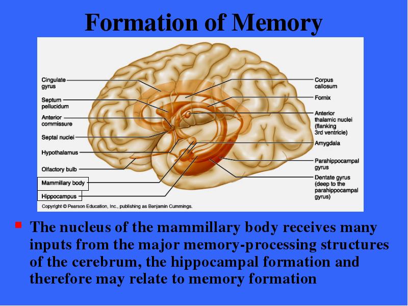
The nucleus of the mammillary body receives many inputs from the major memory-processing structures of the cerebrum, the hippocampal formation and therefore may relate to memory formation
№172 слайд

Содержание слайда: Epithalamus
The epithalamus is the posterior portion of the diencephalon
It forms the roof of the third ventricle
№173 слайд
Содержание слайда: The Epithalamus
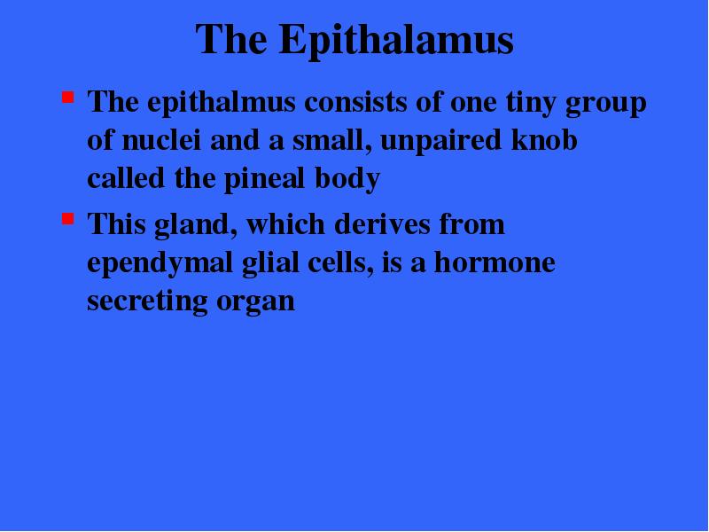
The epithalmus consists of one tiny group of nuclei and a small, unpaired knob called the pineal body
This gland, which derives from ependymal glial cells, is a hormone secreting organ
№174 слайд

Содержание слайда: Epithalamus
The pineal gland extends from the posterior border of the epithalamus
The pineal gland secretes the hormone melatonin which signals the sleep- wake cycle
№175 слайд

Содержание слайда: The Epithalamus
A cerebrospinal fluid-forming structure called a choroid plexus is also part of the epithalamus
№176 слайд

Содержание слайда: The Brain Stem
The third of the four major parts of the brain is the brain stem
From superior to inferior, the brain stem is divided into;
Midbrain
Pons
Medulla oblongata
№177 слайд

Содержание слайда: The Brain Stem
Each region is roughly an inch long
Together than constitute 2.5% of total brain mass
The brain stem has several functions
It produce the rigidly programmed, automatic behaviors necessary for our survival
Acts as a passageway for all the fiber tracts running between the cerebrum and spinal cord
It is heavily involved with the innervation of the face and head as 10 of the 12 cranial nerve attach to it
№178 слайд
Содержание слайда: The Brain Stem
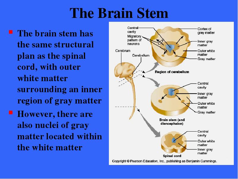
The brain stem has the same structural plan as the spinal cord, with outer white matter surrounding an inner region of gray matter
However, there are also nuclei of gray matter located within the white matter
№179 слайд

Содержание слайда: The Midbrain
The midbrain is located between the diencephalon superiorly and the pons inferiorly
№180 слайд

Содержание слайда: The Midbrain
Its central cavity is the cerebral aqueduct, which divides it into a tectum (dorsal surface) and paired cerebral peduncles
From an anterior view the cerebral peduncles appear as columns that hold up the cerebrum
№181 слайд
Содержание слайда: The Midbrain
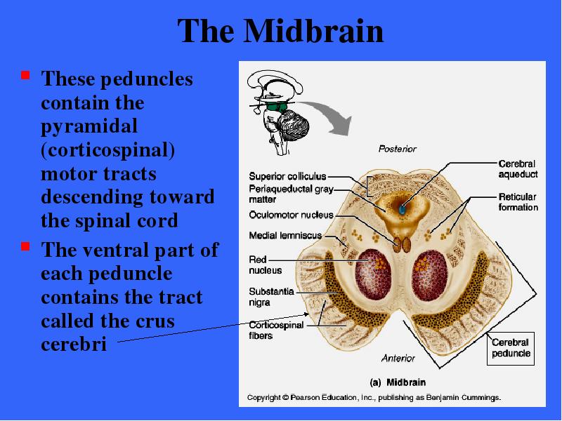
These peduncles contain the pyramidal (corticospinal) motor tracts descending toward the spinal cord
The ventral part of each peduncle contains the tract called the crus cerebri
№182 слайд

Содержание слайда: The Midbrain
Dorsally, the midbrain has the superior cerebellar peduncles which connect midbrain to the cerebellum
№183 слайд

Содержание слайда: The Midbrain
Surrounding the cerebral aqueduct is the peri-aqueductal gray matter that has two somewhat related functions
№184 слайд

Содержание слайда: The Midbrain
The periaqueductal gray matter is involved in the “fright-and-flight” sympathetic reaction
The gray matter is a link between the amygdala of the forebrain (which perceives fear) and the autonomic pathway (which directly signals the physiological reactions associated with fear)
№185 слайд

Содержание слайда: The Midbrain
The gray matter elicits
A terror-induced increase in heart rate
Skyrocketing blood pressure
Wild fleeing or defensive freezing
The flexing of the spine as in curling into a ball for protection
The suppression of pain upon injury
№186 слайд

Содержание слайда: The Midbrain
The periaqueductal gray matter also seems to mediate our response to visceral pain (as when nauseous) during which it
Decreases heart rate and blood pressure
Produce a cold sweat
Discourages movement
№187 слайд
Содержание слайда: The Midbrain
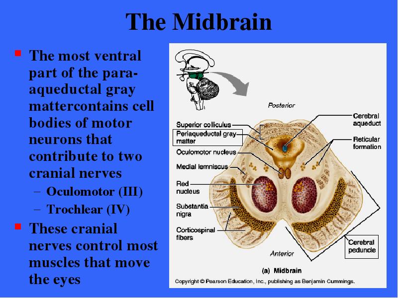
The most ventral part of the para- aqueductal gray mattercontains cell bodies of motor neurons that contribute to two cranial nerves
Oculomotor (III)
Trochlear (IV)
These cranial nerves control most muscles that move the eyes
№188 слайд

Содержание слайда: The Midbrain
Nuclei are also scattered in the surrounding white matter
The largest of these nuclei are the corpora quadrigemia which raise four dome like protrusions on the dorsal midbrain surface
№189 слайд

Содержание слайда: The Midbrain
The superior pair of nuclei, the superior colliculus are visual reflex centers that
coordinate head and eye movements when following a moving object
Make us turn our head involuntarily when we detect movement in our peripherial vision
№190 слайд

Содержание слайда: The Midbrain
The inferior colliculus are part of the auditory relay from the hearing receptors of the ear to the sensory cortex
Act in reflexive response to sound as in the startle reflex
Turn your head toward unexpected source of sound
№191 слайд

Содержание слайда: The Midbrain
Also imbedded in the white matter of the midbrain are two pigmented nuclei, the substantia nigra and the red nucleus
№192 слайд

Содержание слайда: The Midbrain
The substantia nigra is a bandlike nucleus located deep to the cerebral peduncle
It is the largest nuclear mass in the midbrain
№193 слайд

Содержание слайда: The Midbrain
Its dark color reflects its high content of melanin pigment, a precursor of dopamine a neurotransmitter released by these neurons
The substantia nigra is functionally linked to the basal nuclei of the cerebral hemispheres
№194 слайд

Содержание слайда: The Midbrain
The red nucleus is found between the substantia nigra and the cerebral aqueduct
It reddish hue is due to its vascular supply and the presence of iron pigment in the cell bodies of its neurons
№195 слайд
Содержание слайда: The Midbrain
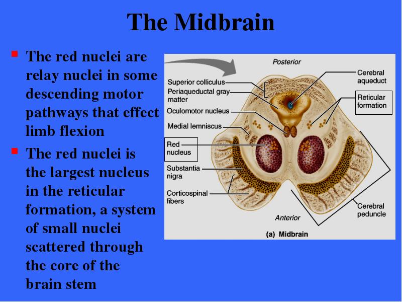
The red nuclei are relay nuclei in some descending motor pathways that effect limb flexion
The red nuclei is the largest nucleus in the reticular formation, a system of small nuclei scattered through the core of the brain stem
№196 слайд

Содержание слайда: The Pons
The pons is the bulging brain stem region wedged between the midbrain and the medulla oblongata
№197 слайд

Содержание слайда: The Pons
It forms part of the anterior wall of the fourth ventricle
It is chiefly composed of conduction tracts
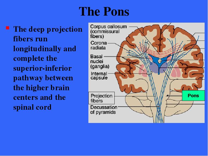
The deep projection fibers run longitudinally and complete the pathway between higher brain centers and spinal cord
№198 слайд
Содержание слайда: The Pons
The deep projection fibers run longitudinally and complete the superior-inferior pathway between the higher brain centers and the spinal cord
№199 слайд

Содержание слайда: The Pons
The more superficial nuclei are relays for conversations between the motor cortex and the cerebellum
These fibers are orientated dorsally and transversely and connect the pons bilaterally with the cerebellum
№200 слайд

Содержание слайда: The Pons
Several cranial nerves issue from pons nuclei
Trigeminal nerve
Abducens nerve
Facial nerves
№201 слайд

Содержание слайда: The Pons
Other important pons nuclei are part of the reticular formation
The pneumotaxic center is a respiratory center
Functioning with medullary respiratory centers it helps to maintain the normal rhythm of breathing
№202 слайд
Содержание слайда: The Medulla Oblongata
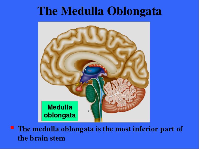
The medulla oblongata is the most inferior part of the brain stem
№203 слайд

Содержание слайда: The Medulla Oblongata
The medulla blends into the spinal cord at the level of the foramen magnum
The central canal of the spinal cord continues upward into the medulla where it broaden to form the fourth ventricle
№204 слайд

Содержание слайда: The Medulla Oblongata
The medulla has several externally visible landmarks which form longitudinal ridges on the ventral surface called the pyramids
These are formed by the large pyramidal tracts descending from the motor cortex
№205 слайд

Содержание слайда: The Medulla Oblongata
Just above the medulla-spinal cord junction most of the fibers cross over to the opposite side before continuing their descent into the spinal cord
The crossover point is called the decussation of the pyramids
№206 слайд

Содержание слайда: The Medulla Oblongata
The consequence of this crossover is that each hemisphere chiefly controls the voluntary movements of muscles on the opposite or contralateral side of the body
№207 слайд

Содержание слайда: The Medulla Oblongata
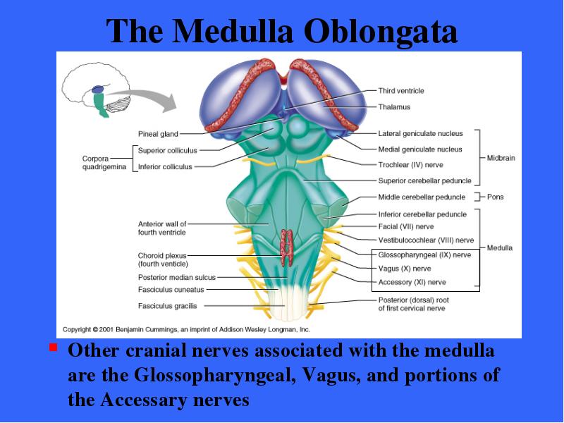
The inferior cerebellar peduncles are fiber tracts that connect the medulla to the cerebellum dorsally
The olives are oval swellings produced by the underlying inferior olivary nuclei
№208 слайд

Содержание слайда: The Medulla Oblongata
The olivary nuclei relay sensory information on the state of stretch of our muscles and joints to the cerebellum
№209 слайд
Содержание слайда: The Medulla Oblongata
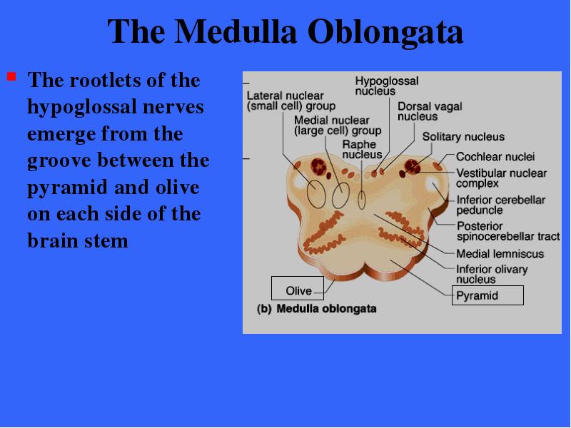
The rootlets of the hypoglossal nerves emerge from the groove between the pyramid and olive on each side of the brain stem
№210 слайд
Содержание слайда: The Medulla Oblongata
Other cranial nerves associated with the medulla are the Glossopharyngeal, Vagus, and portions of the Accessary nerves
№211 слайд

Содержание слайда: The Medulla Oblongata
The fibers of the vestibulocochlear synapse with the cochlear nuclei which receive information on auditory inputs
№212 слайд

Содержание слайда: The Medulla Oblongata
Also housed within the medulla are several nuclei associated with ascending sensory tracts
The most dominant of these are the dorsally located nucleus gracilis and nucleus cuneatus associated with the medial lemniscal tract
№213 слайд

Содержание слайда: Medulla
Oblongata
These serve as relay nuclei in a pathway by which general somatic sensory information ascends from the spinal cord to the somatosensory cortex
№214 слайд

Содержание слайда: The Medulla Oblongata
The medulla has a critical role as an autonomic reflex center involved in maintaining body homeostasis
The cardiovascular center
The respiratory centers
Other centers
№215 слайд

Содержание слайда: The Medulla Oblongata
The cardiac center
The cardiac center adjusts the force and rate of heart contraction to meet bodily needs
№216 слайд

Содержание слайда: The Medulla Oblongata
The vasomotor center
The vasomotor center regulates blood pressure by acting on smooth muscle in the walls of the blood vessels to effect changes in blood vessel diameter
Vasoconstriction causes blood pressure to rise; dilation reduces blood pressure
№217 слайд

Содержание слайда: The Medulla Oblongata
The respiratory centers
The medullary respiratory centers control the rate and depth of breathing and maintains respiratory rhythm
№218 слайд
Содержание слайда: The Medulla Oblongata
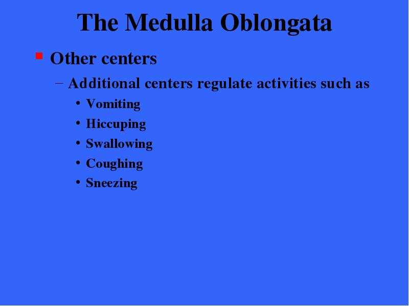
Other centers
Additional centers regulate activities such as
Vomiting
Hiccuping
Swallowing
Coughing
Sneezing
№219 слайд

Содержание слайда: The Medulla Oblongata
Many functions of the medulla overlap with those attributed to the hypothalamus
The overlap is easily explained
The hypothalamus exerts its control over most visceral functions by relaying its instructions through the medulla’s reticular centers (within the Medulla oblongata) which carry them out
№220 слайд

Содержание слайда: The Cerebellum
The cerebellum is exceeded in size only by the cerebrum
It accounts for about 11% of total brain mass
№221 слайд

Содержание слайда: The Cerebellum
The cerebellum is located dorsal to the pons and medulla under the occipital lobe of the cerebral hemispheres
№222 слайд

Содержание слайда: The Cerebellum
It is separated from the occipital lobe by the transverse fissure
It rests in the posterior cranial fossa of the skull
№223 слайд

Содержание слайда: The Cerebellum
The cerebellum processes inputs received from
Cerebral motor cortex
Various brain stem nuclei
Sensory receptors
The cerebellum provides precise timing and appropriate patterns of skeletal muscle contraction
Need for the smooth, coordinated movements of daily living
Cerebeller activity occurs subconsciously; we have no awareness of its functioning
№224 слайд

Содержание слайда: The Cerebellum
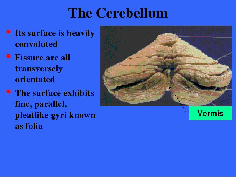
The cerebellum is bilaterally symmetrical
Its two cerebellar hemispheres are connected medially by the wormlike vermis
№225 слайд
Содержание слайда: The Cerebellum
Its surface is heavily convoluted
Fissure are all transversely orientated
The surface exhibits fine, parallel, pleatlike gyri known as folia
№226 слайд

Содержание слайда: The Cerebellum
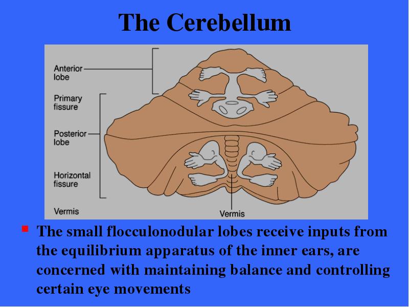
Deep fissures divide each hemisphere into three lobes
Anterior lobe
Posterior lobe
Flocculonodular lobe* (Cannot be seen in a surface view)
№227 слайд

Содержание слайда: The Cerebellum
The cerebellum has a thin outer cortex of gray matter
Internal white matter
Small, deeply situated paired masses of gray matter
№228 слайд
Содержание слайда: The Cerebellum
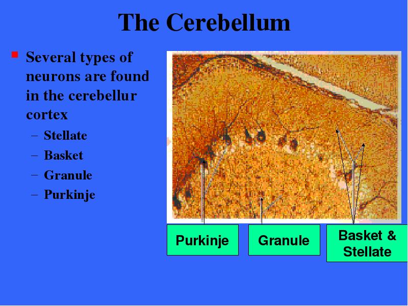
Several types of neurons are found in the cerebellur cortex
Stellate
Basket
Granule
Purkinje
№229 слайд

Содержание слайда: The Cerebellum
The large Purkinje cells with their extensively branched dendrites are the only cortical neurons that send their axons through the white matter to synapse with the central nuclei of the cerebellum
These nuclei mediate most of the output of the cerebellum
№230 слайд

Содержание слайда: The Cerebellum
The anterior and posterior lobes of the cerebellum act to coordinate body movements
The lobes have completely overlapping sensory and motor maps of the entire body
№231 слайд

Содержание слайда: The Cerebellum
The medial portions receive information from the axial portion of the body and influence the motor activities of the trunk and girdle muscles by relaying information to the cerebral motor cortex
№232 слайд

Содержание слайда: The Cerebellum
The intermediate parts of each hemisphere are more concerned with the distal parts of the limbs and skilled movements
№233 слайд

Содержание слайда: The Cerebellum
The lateral parts of each hemisphere receive inputs from the association areas of the cerebral cortex and appear to play a role in planning rather than executing movements
№234 слайд
Содержание слайда: The Cerebellum
The small flocculonodular lobes receive inputs from the equilibrium apparatus of the inner ears, are concerned with maintaining balance and controlling certain eye movements
№235 слайд

Содержание слайда: The Cerebellum
Three pairs of fiber tracts, cerebellur peduncles connect the the cerebellum to the brain stem
№236 слайд

Содержание слайда: The Cerebellum
Virtually all fibers entering and leaving the cerebellum are ipsilateral; from and to the same side of the body
№237 слайд

Содержание слайда: The Cerebellum
The superior cerebellar peduncles connect the cerebellum and the midbrain. Fibers in these peduncles originate in the deep cerebellar nuclei and communicate with the cerebral motor cortex via thalamic relays
№238 слайд

Содержание слайда: The Cerebellum
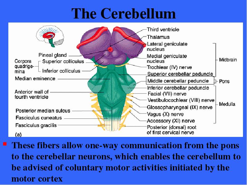
The middle cerebellar peduncles connect the pons the cerebellum.
№239 слайд
Содержание слайда: The Cerebellum
These fibers allow one-way communication from the pons to the cerebellar neurons, which enables the cerebellum to be advised of coluntary motor activities initiated by the motor cortex
№240 слайд

Содержание слайда: The Cerebellum
The inferior cerebellar peduncles connect the cerebellum and the medulla
№241 слайд

Содержание слайда: The Cerebellum
These peduncles contain afferent tracts conveying sensory information to the cerebellum from (1) muscle proprioceptors throughout the body and (2) vestibular nuclei of the brain stem concerned with balance & equil.
№242 слайд

Содержание слайда: Cerebellar Processing - 1
The frontal motor association areas of the cerebral cortex indicates its intents to initiate voluntary muscle contractions
Through collateral fibers of the pyramdial tracts, it notifies the cerebellum of its activity
№243 слайд

Содержание слайда: Cerebellar Processing - 2
At the same time, the cerebellum receives information from the proprioceptors throughout the body
Tension in muscles, tendons, and joint positions
From visual and equilibrium pathways
This information enables the cerebellum to determine where the body is and where it is going
More specifically where the parts of the body are located in space and how are they moving
№244 слайд

Содержание слайда: Cerebellar Processing - 3
The cerebellar cortex assesses this information and calculates the best way to coordinate the force, direction, and extent of muscle contraction
Prevents overshoot
Maintains posture
Ensures smooth, coordinated movements
№245 слайд

Содержание слайда: Cerebellar Processing - 4
Via the superior peduncles, the cerebellum dispatches its “blueprint” for coordination to the cerebral motor cortex which makes appropriate adjustments in its motor plan
Cerebellar fibers also flow to brain stem nuclei, such as the red nuclei of the midbrain, which in turn project to motor neurons of the spinal cord
№246 слайд
Содержание слайда: The Cerebellum
The cerebellum continually compares the higher brain’s intention with the body’s performance and sends out messages to initiate the appropriate measures
In this way, it helps to promote smooth voluntary movements that are precise and economical in terms of muscular effort
№247 слайд

Содержание слайда: The Cerebellum
Cerebellar injury results in the loss of muscle tone and clumsy, unsure movements, and sometimes even impaired thoughts about movements
№248 слайд

Содержание слайда: Functional Brain Systems
Functional brain systems are networks of neurons that work together but span relatively large distances with the brain
They are not localized to a specific region of the brain
The Limbic System (distributed within forebrain)
The Reticular Formation (distributed within the brainstem)
№249 слайд

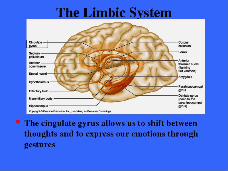
Содержание слайда: The Limbic System
The limbic system is a group of structures located on the medial aspect of each cerebral hemisphere and diencephalon
№250 слайд

Содержание слайда: The Limbic System
The limbic system encircles the upper part of the brain stem and includes
Septal nuclei, Cingulate gyrus, hippocampal formation, and part of the Amygdala,
In the diencephalon the limbic system structures are the hypothalamus and the anterior thalamic nuclei of the thalamus
The fornix and other fiber tracts link these limbic system regions together
№251 слайд
Содержание слайда: The Limbic System
The observation that odors evoke emotional reactions and memories reflects the fact that these structures are linked to the rhinencephalon
№252 слайд

Содержание слайда: The Limbic System
The limbic system is our emotional or affective brain
Two parts seem especially important in emotions
The amygdala
The cingulate gyrus
№253 слайд

Содержание слайда: The Limbic System
The amygdala contains the key nuclei for processing fear and then stimulating the appropriate sympathetic response to fear
№254 слайд

Содержание слайда: The Limbic System
The amygdala also enables us to recognize menacing facial expression in others and to detect the precise direction of the gaze of someone who is looking at us
№255 слайд
Содержание слайда: The Limbic System
The cingulate gyrus allows us to shift between thoughts and to express our emotions through gestures
№256 слайд

Содержание слайда: The Limbic System
The anterior part of the gyrus interprets pain as unpleasant and resolves mental conflict during frustrating tasks
№257 слайд
Содержание слайда: The Limbic System
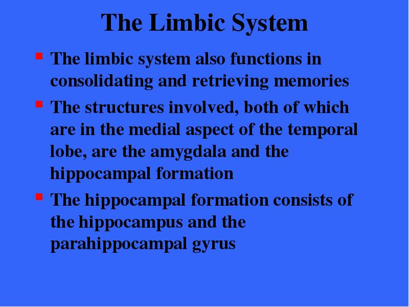
The limbic system also functions in consolidating and retrieving memories
The structures involved, both of which are in the medial aspect of the temporal lobe, are the amygdala and the hippocampal formation
The hippocampal formation consists of the hippocampus and the parahippocampal gyrus
№258 слайд

Содержание слайда: The Limbic System
The hippocampal formation encodes, consolidates, and later retrieves memories of facts and events
It first receives information to be remembered from the rest of the cerebral cortex; then it processes these data and returns then to the cortex, where they are stored as long-term memories
№259 слайд

Содержание слайда: The Limbic System
The amygdala forms memories of experiences that are based entirely on their emotional impact, especially if related to fear
If we later are reminded of these experiences, the amygdala retreives the memories and causes us to re-experience the original emotion
The benefit is that it lets us make difficult and risky decisions correctly, based on memories of our past emotional experiences
№260 слайд

Содержание слайда: The Limbic System
The limbic system communicates with many other regions of the brain
Most output from the limbic system is relayed through the hypothalamus and the reticular formation, the portions of our brain that control our visceral responses
№261 слайд

Содержание слайда: The Limbic system
This fact explains why people under emotional stress experience visceral illnesses such as high blood pressure and heartburn
The limbic system also interacts heavily with the prefrontal lobes of the cerebral cortex
Thus, our feelings (mediated by the emotional brain) and our thoughts (mediated by the thinking brain) interact closely
№262 слайд

Содержание слайда: The Limbic System
We react emotionally to things we consciously understand to be happening
We are consciously aware of the emotional aspect of our lives
№263 слайд

Содержание слайда: The Limbic System
Communication between the cerebral cortex and the limbic system explains why emotions sometimes override logic
It also explains why reason can stop us from expressing our emotions in inappropriate ways
№264 слайд

Содержание слайда: The Reticular Formation
The reticular formation extends through the central core of the medulla oblongata, pons, and midbrain
№265 слайд

Содержание слайда: The Reticular Formation
It is an intricate system composed of loosely clustered neurons in what is otherwise white matter
№266 слайд

Содержание слайда: The Reticular Formation
Reticular neurons can be localized into three broad columns along the length of the brain stem
Raphe
Medial nuclear (large cell) group
Lateral nuclear (small cell) group
№267 слайд
Содержание слайда: The Reticular Formation
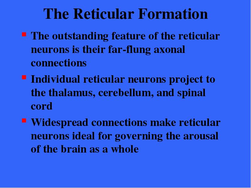
The outstanding feature of the reticular neurons is their far-flung axonal connections
Individual reticular neurons project to the thalamus, cerebellum, and spinal cord
Widespread connections make reticular neurons ideal for governing the arousal of the brain as a whole
№268 слайд

Содержание слайда: The Reticular Formation
Certain reticular neurons send a continuous stream of impulses to the cerebrum (through relays in the thalamus) thereby maintaining the cerebral cortex in an alert state
This arm of the reticular formation is called the reticular activating system or RAS
№269 слайд

Содержание слайда: The Reticular Activating System
The RAS synapses with all major ascending sensory tracts enhancing arousal of the cerebrum
The RAS functions in sleep and in arousal from sleep
It can be affected by general anesthesia, alcohol, tranquilizers, and sleep inducing drugs
Head trauma can also lead to loss of consciousness
№270 слайд

Содержание слайда: Reticular Formation
The RAS also acts as a filter to dampen repetitive, familiar, or weak signals
It is estimated that 99% of all sensory stimuli is disregarded as unimportant
№271 слайд

Содержание слайда: The Reticular Activating System
The activity of the RAS is inhibited by sleep centers in the hypothalamus and other neural regions
Damage to the RAS limits arousal and can result in coma
№272 слайд

Содержание слайда: The Reticular Formation
The reticular formation also has a motor component
Some if its motor nuclei project to motor neurons in the spinal cord via the reticulospinal tracts
These help control the skeletal muscles during coarse movements of the limbs
Other reticular motor nuclei are autonomic centers that regular visceral motor functions (heart rate & respiration)
№273 слайд
Содержание слайда: Protection of the Brain
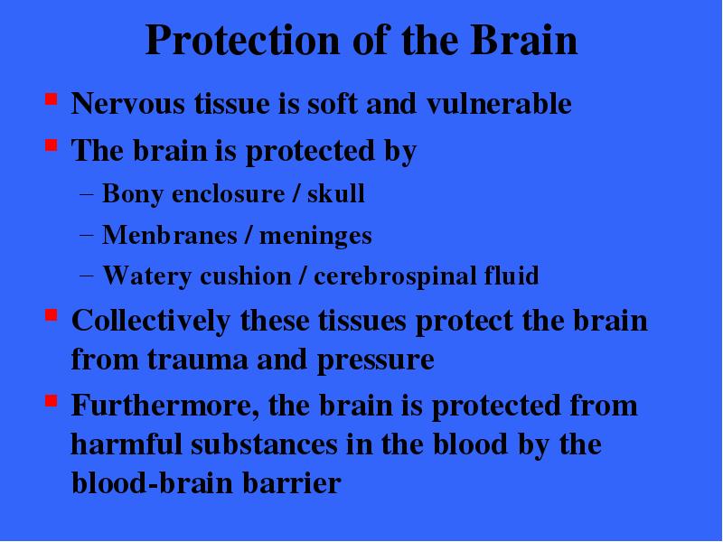
Nervous tissue is soft and vulnerable
The brain is protected by
Bony enclosure / skull
Menbranes / meninges
Watery cushion / cerebrospinal fluid
Collectively these tissues protect the brain from trauma and pressure
Furthermore, the brain is protected from harmful substances in the blood by the blood-brain barrier
№274 слайд

Содержание слайда: Meninges
The meninges are three connective tissue membranes that lie just external to the central nervous system organs
№275 слайд

Содержание слайда: Meninges
The meningeal membranes
Cover and protect the CNS organs
Protect blood vessels and enclose venous sinuses
Contain cerebrospinal fluid
Form partitions within the skull
№276 слайд

Содержание слайда: Meninges
The meninges are three connective tissue membranes that lie just external to the central nervous system organs
№277 слайд

Содержание слайда: Meninges
From external to internal, the meningeal layers are
Dura mater
Arachnoid
Pia mater
№278 слайд

Содержание слайда: The Dura Mater
The leathery dura mater is by far the strongest of the meninges
Where it surrounds the brain it is a double layer membrane
№279 слайд
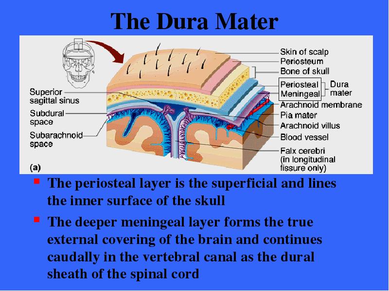
Содержание слайда: The Dura Mater
The periosteal layer is the superficial and lines the inner surface of the skull
The deeper meningeal layer forms the true external covering of the brain and continues caudally in the vertebral canal as the dural sheath of the spinal cord
№280 слайд

Содержание слайда: The Dura Mater
The brain’s dural layers are fused together except in certain areas where they enclose the dural sinuses
The dural sinuses collect venous blood and direct it into the internal jugular veins of the neck
№281 слайд
Содержание слайда: The Dura Mater
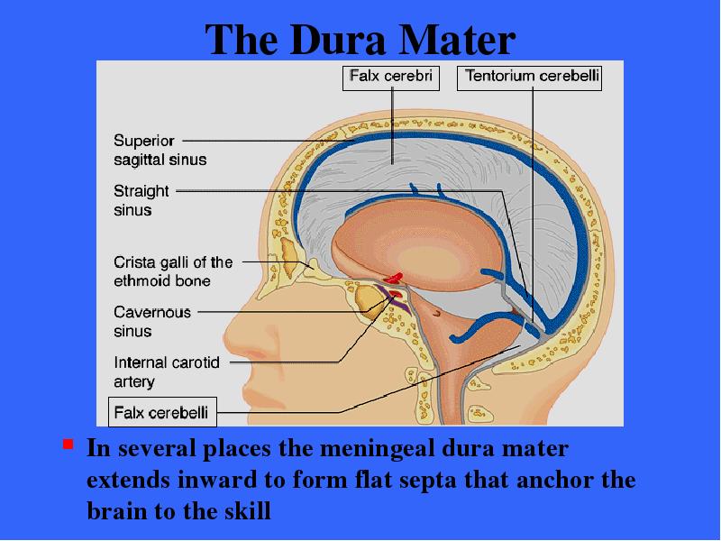
In several places the meningeal dura mater extends inward to form flat septa that anchor the brain to the skill
№282 слайд

Содержание слайда: The Dura Mater
The falx cerebri dips into the longitudinal fissue
It attaches to the crista galli of the ethmoid bone
№283 слайд

Содержание слайда: The Dura Mater
The falx cerebelli forms a midline partition that runs along the vermis of the cerebellum
№284 слайд

Содержание слайда: The Dura Mater
The tentorium cerebelli extends into the transverse fissue between the cerebral hemispheres and the cerebellum
№285 слайд

Содержание слайда: The Arachnoid Mater
The middle membrane forms a loose brain covering over the surface of the cerebrum
It is separated from the dura mater by a narrow serous cavity, the subdural space
Beneath the arachnoid membrane is the wide subarachnoid space
№286 слайд

Содержание слайда: The Arachnoid Mater
The subarachnoid space is filled with cerebrospinal fluid and contains the largest blood vessels serving the brain
Since the arachnoid is fine and elastic, these blood vessels are rather poorly protected
№287 слайд

Содержание слайда: The Arachnoid Mater
Arachnoid villi protrude through the overlying dura mater and into the dural sinuses overlying the superior aspect of the brain
Cerebrospinal fluid is absorbed into the venous blood sinuses through these valvelike villi
№288 слайд
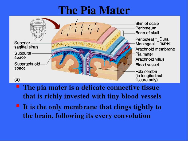
Содержание слайда: The Pia Mater
The pia mater is a delicate connective tissue that is richly invested with tiny blood vessels
It is the only membrane that clings tightly to the brain, following its every convolution
№289 слайд

Содержание слайда: Cerebrospinal Fluid (CSF)
CSF is found in and around the brain and spinal cord
It forms a liquid cushion that gives bouyancy to the CNS organs
By floating the brain, the CPF reduces brain weight by 97% and thus prevents the brain from crushing under its own weight
CSF also protects the brain and spinal cord from trauma
CSF also helps to nourish the brain
№290 слайд
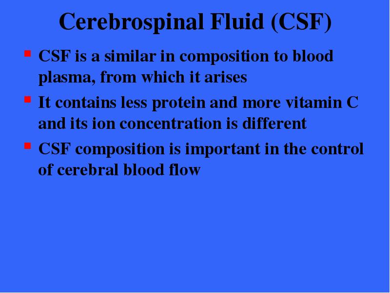
Содержание слайда: Cerebrospinal Fluid (CSF)
CSF is a similar in composition to blood plasma, from which it arises
It contains less protein and more vitamin C and its ion concentration is different
CSF composition is important in the control of cerebral blood flow
№291 слайд

Содержание слайда: Choroid Plexus
Choroid plexus hang from the roof of each ventricle
These plexuses form CSF
The plexuses are clusters of thin walled capillaries enclosed by a layer of ependymal cells
№292 слайд

Содержание слайда: Choroid Plexus
The capillaries of the choroid plexus are fairly permeable and tissue fluid filter continuously from the bloodstream
№293 слайд

Содержание слайда: Choroid Plexus
The choroid plexus cells are joined by tight junctions and have ion pumps that allow them to modify this filtrate by actively transporting only certain ions across their membranes into the CSF pool
№294 слайд

Содержание слайда: The Choroid Plexus
In adults, the total CSF volume of about 150 ml is replaced every 3-4 hours
900 ml is produced daily
The choroid plexus also helps to cleanse the CSF by removing waste products and other unnecessary solutes
Once produced CSF moves freely through the ventricles
№295 слайд
Содержание слайда: CSF Circulation
Most CSF enters the subarachnoid space via the apertures in the walls of the fourth ventricle
The motion of the CSF is aided by the long microvilli of the ependymal cells lining the ventricles
Some CSF enters the central canal of the spinal cord
№296 слайд

Содержание слайда: CSF Circulation
In the subarachnoid space the CSF bathes the outer surface of the brain and cord and then returns to the blood in the dural sinuses via the arachnoid villi
№297 слайд
Содержание слайда: Blood-Brain Barrier
The barrier is a protective mechanism that helps maintain a stable environment for the brain
The brain is very dependent on a contant internal environment
Fluctuations in the concentration of ions, hormones, or amino acids, would alter the brain’s function
Hormones and amion acids / neurotransmitters
Ions / neuron thresholds (K+)
№298 слайд

Содержание слайда: Blood-Brain Barrier
Bloodborne substances within the brain’s capillaries are separated from the extra- cellular space and neurons by
Continuous endothelium of the capillary walls
Relatively thick basal lamina surrounding the external face of the capillary
To a limited extend the “feet” of the astrocytes that cling to the capillaries
№299 слайд

Содержание слайда: Blood-Brain Barrier
The capillary endothelial cells are joined almost seamlessly by tight junctions
They are the least permeable capillaries in the body
The relative impermeability of brain capillaries accounts for most of the blood brain barrier
№300 слайд
Содержание слайда: Blood-Brain Barrier
The blood-brain barrier is a selective, rather than absolute barrier
Nutrients, such as glucose, essential amino acids, and some electrolytes, move passively by facilitated diffusion through the endothelial cell membranes
Bloodborne metabolic wastes, such as urea and creatinine as well as proteins, certain toxins, and most drugs, are prevented from entering brain tissue
№301 слайд

Содержание слайда: Blood-Brain Barrier
The barrier is ineffective against fats, fatty acids, oxygen, and carbon dioxide, and other fat-soluble molecules that diffuse easily through all plasma membranes
This explains why bloodborne alcohol, nicotine, and anesthetics can affect the brain
The barrier is not completely uniform and not completely developed in infants
№302 слайд

Содержание слайда: Homeostatic Imbalances
Traumatic Brain Injuries
Concussion / Contusion
Cerebrovascular Accidents
Strokes / Transient ischemic attacks
Degenerative Brain Diseases
Alzheimer’s Disease / Ach deficits
Parkinson’s Disease / Degeneration of the substantia nigra
Huntington’s Disease / Degeneration of the basal nuclei
№303 слайд

Содержание слайда: The Brain
End of this section
Chapter 12 Continues
Скачать все slide презентации Central Nervous System одним архивом: