Презентация Data Collection of Primary Central Nervous System (CNS) Tumors онлайн
На нашем сайте вы можете скачать и просмотреть онлайн доклад-презентацию на тему Data Collection of Primary Central Nervous System (CNS) Tumors абсолютно бесплатно. Урок-презентация на эту тему содержит всего 157 слайдов. Все материалы созданы в программе PowerPoint и имеют формат ppt или же pptx. Материалы и темы для презентаций взяты из открытых источников и загружены их авторами, за качество и достоверность информации в них администрация сайта не отвечает, все права принадлежат их создателям. Если вы нашли то, что искали, отблагодарите авторов - поделитесь ссылкой в социальных сетях, а наш сайт добавьте в закладки.
Оцените презентацию от 1 до 5 баллов!
- Тип файла:ppt / pptx (powerpoint)
- Всего слайдов:157 слайдов
- Для класса:1,2,3,4,5,6,7,8,9,10,11
- Размер файла:1.89 MB
- Просмотров:66
- Скачиваний:0
- Автор:неизвестен
Слайды и текст к этой презентации:
№1 слайд
Содержание слайда: Data Collection of Primary Central Nervous System (CNS) Tumors
№2 слайд
Содержание слайда: Portions of this presentation are based on non-malignant CNS tumor data collection rules adopted by the North American Association of Central Cancer Registries (NAACCR) Uniform Data Standards Committee - June 2003.
Portions of this presentation are based on non-malignant CNS tumor data collection rules adopted by the North American Association of Central Cancer Registries (NAACCR) Uniform Data Standards Committee - June 2003.
№3 слайд
Содержание слайда: Part I
Rationale
History
Definition of Reportable Cases
Casefinding
Anticipated Impact on Registries
№4 слайд
Содержание слайда: Rationale for Non-malignant CNS Tumor Surveillance and Registration
Non-malignant CNS tumors cause disruption in normal function similar to that caused by malignant CNS tumors.
Location of a CNS tumor is as important as tumor behavior (benign or malignant) to morbidity and mortality.
№5 слайд
Содержание слайда: History 1992 -1996
1992 Central Brain Tumor Registry of the United States (CBTRUS) formed to report population-based data on primary benign, borderline, and malignant CNS tumors.
1996 National Coordinating Council on Cancer Surveillance (NCCCS) formed Brain Tumor Working Group (BTWG) to explore the feasibility of registering non-malignant CNS tumors
№6 слайд

Содержание слайда: History 1998
BTWG forwarded four recommendations to the NCCCS
NCCCS
Accepted recommendations 1 and 2
Deferred recommendations 3 and 4
№7 слайд
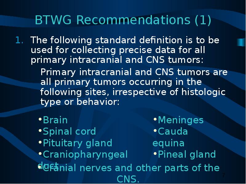
Содержание слайда: BTWG Recommendations (1)
The following standard definition is to be used for collecting precise data for all primary intracranial and CNS tumors:
Primary intracranial and CNS tumors are all primary tumors occurring in the following sites, irrespective of histologic type or behavior:
№8 слайд
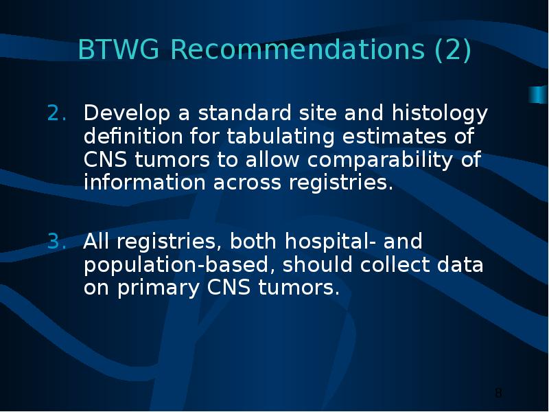
Содержание слайда: BTWG Recommendations (2)
Develop a standard site and histology definition for tabulating estimates of CNS tumors to allow comparability of information across registries.
All registries, both hospital- and population-based, should collect data on primary CNS tumors.
№9 слайд

Содержание слайда: BTWG Recommendations (3)
Develop training for reporting and tabulating primary intracranial and CNS tumors, and develop computerized edit- checking procedures.
№10 слайд

Содержание слайда: History 2000
International Classification of Diseases for Oncology 3rd Edition (ICD-O-3) and World Health Organization (WHO) 2000 Brain Tumor Classification are compatible.
November
Consensus conference on brain tumor definition convened. Group agrees to:
Site definition as in Recommendation 1.
Need to develop a standard site and histology definition based on the SEER site and histology validation list.
№11 слайд

Содержание слайда: History 2001-2002
2001 NCCCS
Accepted Recommendations 1 and 2 as completed.
Reconvened the BTWG to work on Recommendations 3 and 4.
2002 NAACCR established subcommittee of Registry Operations Committee to:
Identify changes needed in registry operations for inclusion of non-malignant CNS tumors.
October: Benign Brain Tumor Cancer Registries Amendment Act (Public Law 107-260) signed by President Bush.
№12 слайд

Содержание слайда: Reportable Brain-Related Tumors (1)
Public Law 107-260 requires reporting of brain-related tumors.
The term “brain-related tumor” means a listed primary tumor (whether malignant or benign) occurring in any of the following sites:
(I) The brain, meninges, spinal cord, cauda equina, a cranial nerve or nerves, or any other part of the CNS.
(II) The pituitary gland, pineal gland, or craniopharyngeal duct.
№13 слайд

Содержание слайда: Reportable Brain-Related Tumors (2)
Brain
Cerebrum (C71.0)
Frontal lobe (C71.1)
Temporal lobe (C71.2)
Parietal lobe (C71.3)
Occipital lobe (C71.4).
№14 слайд

Содержание слайда: Reportable Brain-Related Tumors (3)
Brain (continued)
Ventricle (C71.5)
Cerebellum (C71.6)
Brain stem (C71.7)
Overlapping lesion of the brain (C71.8)
Brain NOS (C71.9)
№15 слайд
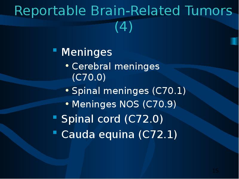
Содержание слайда: Reportable Brain-Related Tumors (4)
Meninges
Cerebral meninges (C70.0)
Spinal meninges (C70.1)
Meninges NOS (C70.9)
Spinal cord (C72.0)
Cauda equina (C72.1)
№16 слайд

Содержание слайда: Reportable Brain-Related Tumors (5)
Cranial nerves
Olfactory nerve (C72.2)
Optic nerve (C72.3)
Acoustic nerve (C72.4)
Cranial nerve NOS (C72.5)
№17 слайд

Содержание слайда: Reportable Brain-Related Tumors (6)
Other CNS (C72.8, C72.9)
Pituitary gland (C75.1)
Craniopharyngeal duct (C75.2)
Pineal gland (C75.3)
For the sites described, benign, borderline, and malignant tumors are reportable for cases diagnosed on or after January 1, 2004.
№18 слайд

Содержание слайда: History 2003
2003 SEER-supported registries and COC-approved hospital cancer registries will also report non-malignant CNS tumors diagnosed on or after January 1, 2004.
№19 слайд
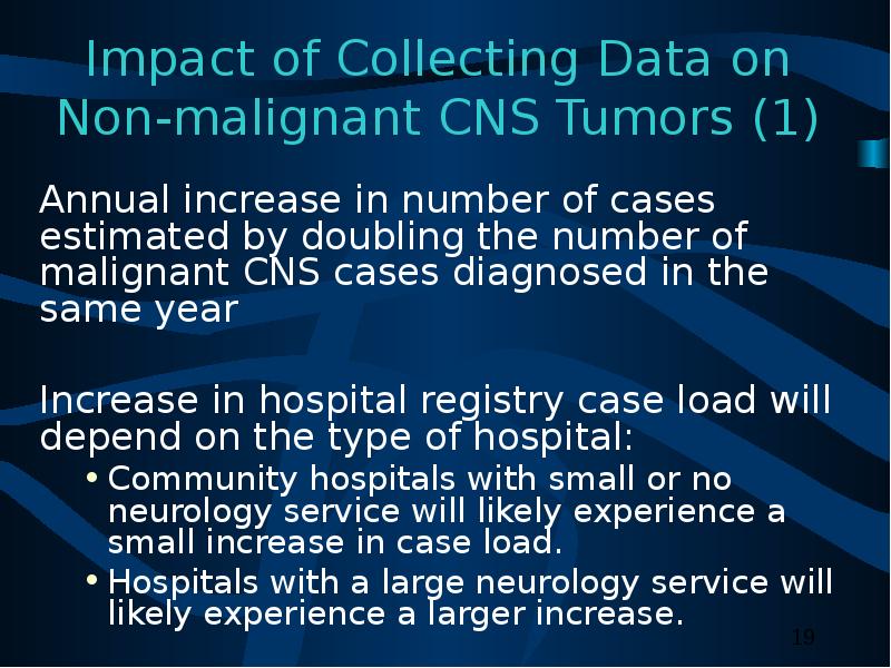
Содержание слайда: Impact of Collecting Data on Non-malignant CNS Tumors (1)
Annual increase in number of cases estimated by doubling the number of malignant CNS cases diagnosed in the same year
Increase in hospital registry case load will depend on the type of hospital:
Community hospitals with small or no neurology service will likely experience a small increase in case load.
Hospitals with a large neurology service will likely experience a larger increase.
№20 слайд

Содержание слайда: Impact of Collecting Data on Non-malignant CNS Tumors (2)
Central registry case load is estimated to increase by 1%.
In 2002, 21 state cancer registries collected data on non-malignant CNS tumors:
Minimal impact if registry’s definition for brain-related sites does not change.
№21 слайд

Содержание слайда: Impact of Collecting Data on Non-malignant CNS Tumors (3)
Central registries adding non-malignant CNS tumors to reportable case definition may have to change state reporting law if law does not allow for collection of data on non-malignant cases.
№22 слайд

Содержание слайда: Impact of Collecting Data on Non-malignant CNS Tumors (4)
All cancer registries must:
Have the same definition for brain-related tumors.
Implement data edits created for non-malignant CNS tumors.
Report rates for these tumors.
№23 слайд

Содержание слайда: Case-finding (1)
Additional or expanded case-finding mechanisms:
Pathology
Radiology
Treatment facilities:
Radiation oncology centers and departments
Gamma or cyber knife center.
№24 слайд

Содержание слайда: Case-finding (2)
Disease indices
Surgery logs
Diagnostic imaging
Radiation oncology
Neurology clinics
Medical oncology
Autopsy reports.
№25 слайд

Содержание слайда: Case-finding Sources
Free-standing radiation therapy centers
Free-standing Magnetic Resonance Imaging (MRI) centers
Free-standing gamma or cyber knife centers
Free-standing oncology centers
Data exchange with other central registries
Death clearance process
№26 слайд

Содержание слайда: ICD-9-CM Codes for Case-finding
№27 слайд
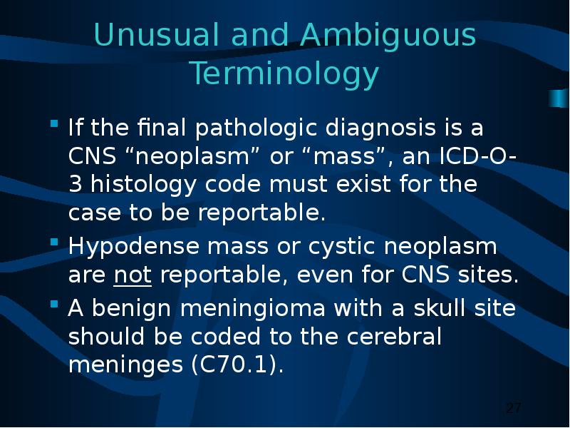
Содержание слайда: Unusual and Ambiguous Terminology
If the final pathologic diagnosis is a CNS “neoplasm” or “mass”, an ICD-O-3 histology code must exist for the case to be reportable.
Hypodense mass or cystic neoplasm are not reportable, even for CNS sites.
A benign meningioma with a skull site should be coded to the cerebral meninges (C70.1).
№28 слайд

Содержание слайда: Part II
CNS Anatomy and Function
Histologies and Primary Sites
Grading Systems and Coding Grade
№29 слайд

Содержание слайда: CNS Functional Anatomy
№30 слайд

Содержание слайда: CNS Anatomy
№31 слайд
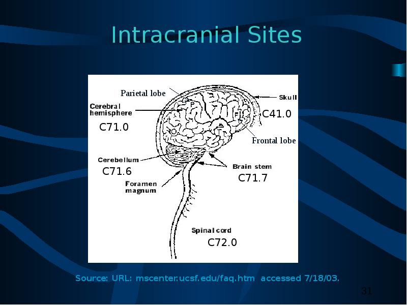
Содержание слайда: Intracranial Sites
№32 слайд
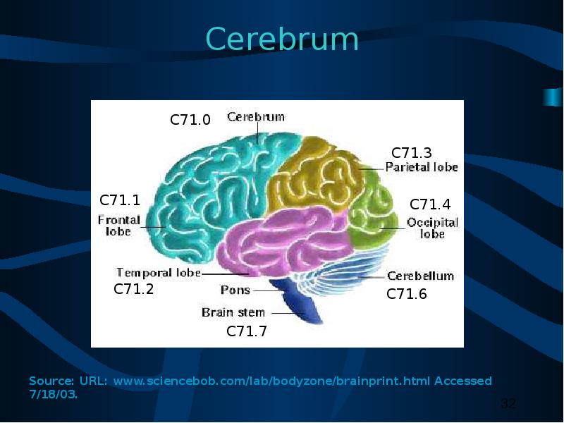
Содержание слайда: Cerebrum
№33 слайд

Содержание слайда: Cerebellum and Brain Stem
№34 слайд
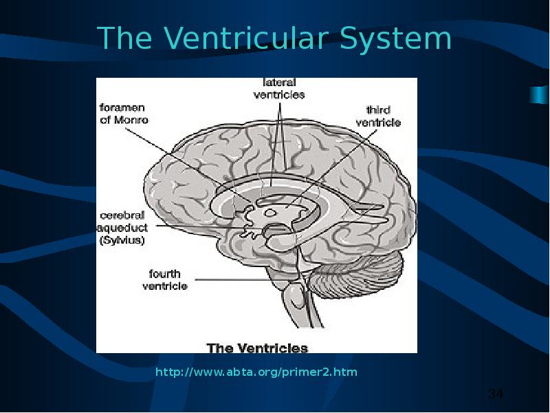
Содержание слайда: The Ventricular System
№35 слайд

Содержание слайда: Pineal and Pituitary Glands
№36 слайд
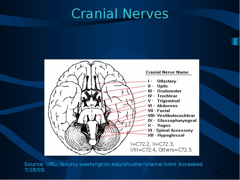
Содержание слайда: Cranial Nerves
№37 слайд

Содержание слайда: Meninges
№38 слайд

Содержание слайда: Tentorium
№39 слайд
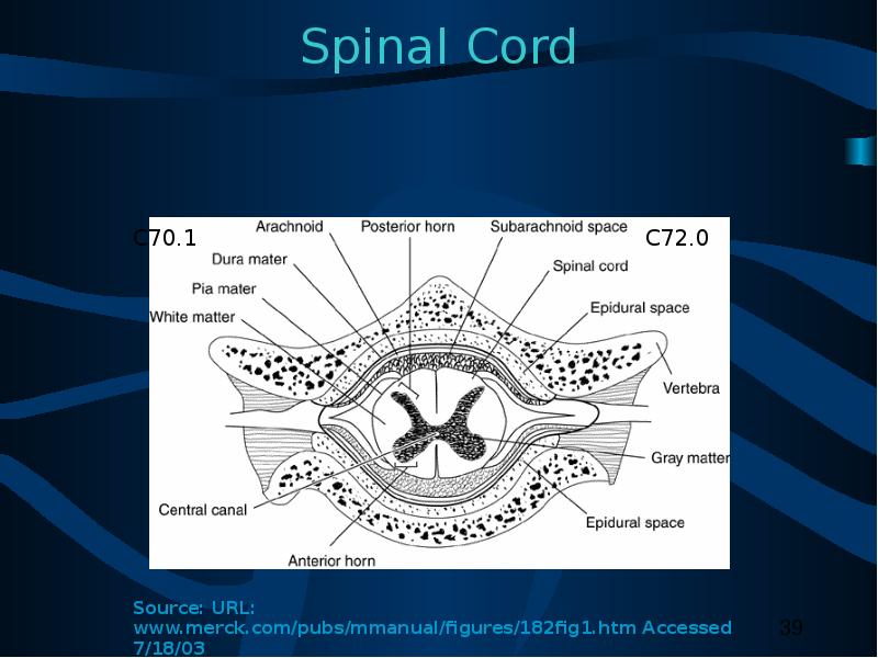
Содержание слайда: Spinal Cord
№40 слайд

Содержание слайда: Cellular Classification
Neuroepithelial tumors
Astrocytomas
Oligodendrogliomas
Ependymomas
Pineal parenchymal tumors
Other CNS tumors
Sellar tumors
Hematopoetic tumors
Germ cell tumors
Meningiomas
Tumors of cranial nerves
№41 слайд

Содержание слайда: Glial Tumors (1)
Glial tissue: supportive tissue of brain made up of astrocytes and oligodendrocytes
Glial tumors assigned ICD-O-3 histology codes from glioma series:
Codes 938 through 948.
№42 слайд

Содержание слайда: Glial Tumors (2)
Astrocytic tumors
Noninfiltrating
Juvenile pilocytic (M9421)
Subependymal (M9383)
Infiltrating
Well-differentiated mildly and moderately anaplastic astrocytomas (M9401)
Anaplastic astrocytomas
Glioblastoma multiforme (M9440)
Brain stem gliomas (M9380)
№43 слайд

Содержание слайда: Glial Tumors (3)
Ependymal tumors
Myxopapillary and well-differentiated ependymomas (M9394)
Anaplastic ependymomas (M9392)
Ependymoblastomas (M9392)
Oligodendroglial tumors
Well-differentiated oligodendrogliomas (M9450)
Anaplastic oligodendrogliomas (M9451)
№44 слайд
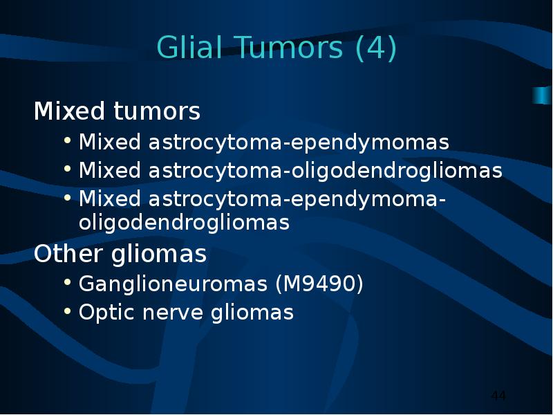
Содержание слайда: Glial Tumors (4)
Mixed tumors
Mixed astrocytoma-ependymomas
Mixed astrocytoma-oligodendrogliomas
Mixed astrocytoma-ependymoma-oligodendrogliomas
Other gliomas
Ganglioneuromas (M9490)
Optic nerve gliomas
№45 слайд

Содержание слайда: Non-Glial Tumors (1)
Pineal region tumors
Parenchymal tumors
Pineocytomas (M9361)
Pineoblastomas (M9362)
Pineal astrocytomas (M9400)
Germ cell tumors
Germinomas (M9064)
Embryonal carcinomas (M9070)
Choriocarcinomas (M9100)
Teratomas (M9080)
№46 слайд

Содержание слайда: Non-Glial Tumors (2)
Meningiomas
Meningioma: Benign (M953_)
Malignant meningiomas
Anaplastic meningioma
Hemangiopericytoma (M9150)
Papillary meningioma (M9538)
Choroid plexus tumors
Choroid plexus papilloma (M9390)
Choroid plexus carcinoma
Choroid plexus meningioma (M9538)
№47 слайд
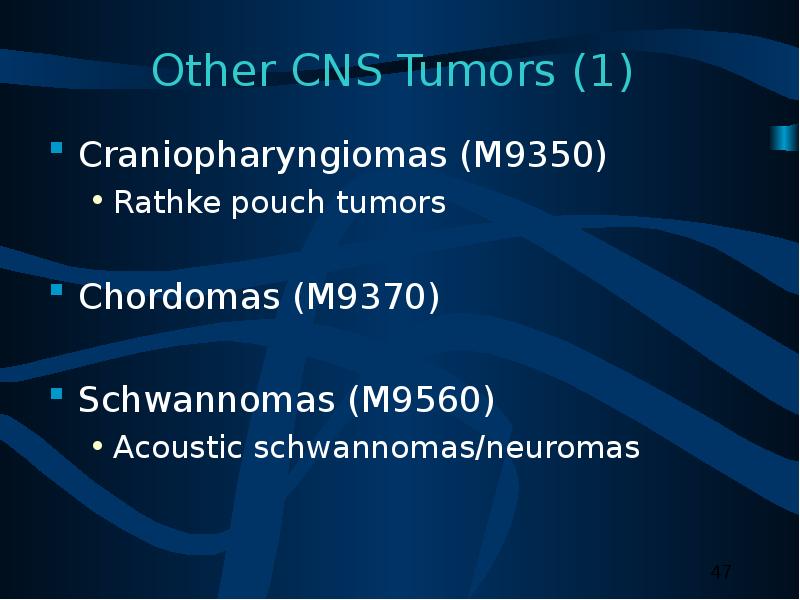
Содержание слайда: Other CNS Tumors (1)
Craniopharyngiomas (M9350)
Rathke pouch tumors
Chordomas (M9370)
Schwannomas (M9560)
Acoustic schwannomas/neuromas
№48 слайд

Содержание слайда: Other CNS Tumors (2)
Embryonal tumors
Retinoblastomas (M9510)
Primitive neuroectodermal tumors (PNETs)
Meduloblastomas (M9470)
Neuroblastomas (M9500)
№49 слайд

Содержание слайда: Other CNS Tumors (3)
Lymphomas (M9590)
Arise from
Indigenous brain histiocytes (microglia)
Rare lymphocytes in meninges
High incidence in patients with AIDS
Vascular tumors
Rare, non-malignant tumors
Arise from blood vessels of brain and spinal cord
Hemangioblastoma (M9161) most common vascular tumor
№50 слайд
Содержание слайда: Other CNS Tumors (4)
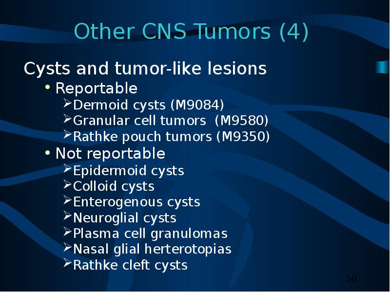
Cysts and tumor-like lesions
Reportable
Dermoid cysts (M9084)
Granular cell tumors (M9580)
Rathke pouch tumors (M9350)
Not reportable
Epidermoid cysts
Colloid cysts
Enterogenous cysts
Neuroglial cysts
Plasma cell granulomas
Nasal glial herterotopias
Rathke cleft cysts
№51 слайд

Содержание слайда: Childhood versus Adult Tumors
CNS tumor histology and location are different in adult and children.
Tumor location and extent of spread affect treatment and prognosis.
Most common solid tumor in childhood.
№52 слайд

Содержание слайда: Childhood Brain Tumors
Meduloblastomas are the most common CNS histology in children.
50% are infratentorial.
Common infratentorial tumors:
Cerebellar astrocytomas
Meduloblastomas
Ependymomas
Brain stem gliomas
Atypical teratoid tumors
№53 слайд

Содержание слайда: Cellular Classification
Childhood Brain Tumors (1)
Supratentorial tumors in children
№54 слайд

Содержание слайда: Cellular Classification
Childhood Brain Tumors (2)
The histopathology of childhood spinal tumors is the same as for childhood brain tumors.
Primary spinal cord tumors comprise approximately 1% to 2% of all childhood CNS tumors.
№55 слайд
Содержание слайда: Cellular Classification
Childhood CNS Tumors
Cause of childhood CNS tumors remains unknown.
American Academy of Pediatrics has outlined guidelines for pediatric cancer centers and their role in the treatment of pediatric cancer patients.
№56 слайд

Содержание слайда: ICD-O-3 Coding Issues (1)
Some histologies may be difficult to determine if the primary site is intracranial or the skull (C41.0).
Non-malignant tumors of the skull are not reportable.
Chondroma (M9220/0) must originate in a brain-related site to be reportable.
Chordoma (M9370/3) and chondrosarcoma (M9220/3) are malignant.
Tumors in brain-related sites are analyzed separately from those in the skull.
№57 слайд
Содержание слайда: ICD-O-3 Coding Issues (2)
Continue to assign histology code M9421/3 to pilocytic astrocytoma.
When the primary site for intracranial schwannoma (9560/0) is not documented in source documents, the site should be coded to cranial nerves NOS (C72.5).
№58 слайд
Содержание слайда: Grade for CNS Tumors
Sixth digit of ICD-O-3 histology code
Describes tumor differentiation or grade.
Is not usually specified for CNS tumors.
Is always assigned code 9 for non-malignant CNS tumors:
Not determined, not stated, or not applicable.
Per ICD-O-3, page 30, Rule G, paragraph 1 “Only malignant tumors are graded.”
Not the same as WHO grade.
№59 слайд

Содержание слайда: WHO Grade (1)
WHO grade coded in Collaborative Stage data field:
Site-specific factor 1 for Brain.
Four-category tumor grading system
Grade I
Slow growing
Non-malignant tumors
Patients have long-term survival.
№60 слайд

Содержание слайда: WHO Grade (2)
Grade II
Relatively slow growing
Sometimes recur as higher grade tumors
May be non-malignant or malignant .
Grade III
Malignant tumors
Often recur as higher grade tumors.
Grade IV
Highly malignant and aggressive.
№61 слайд
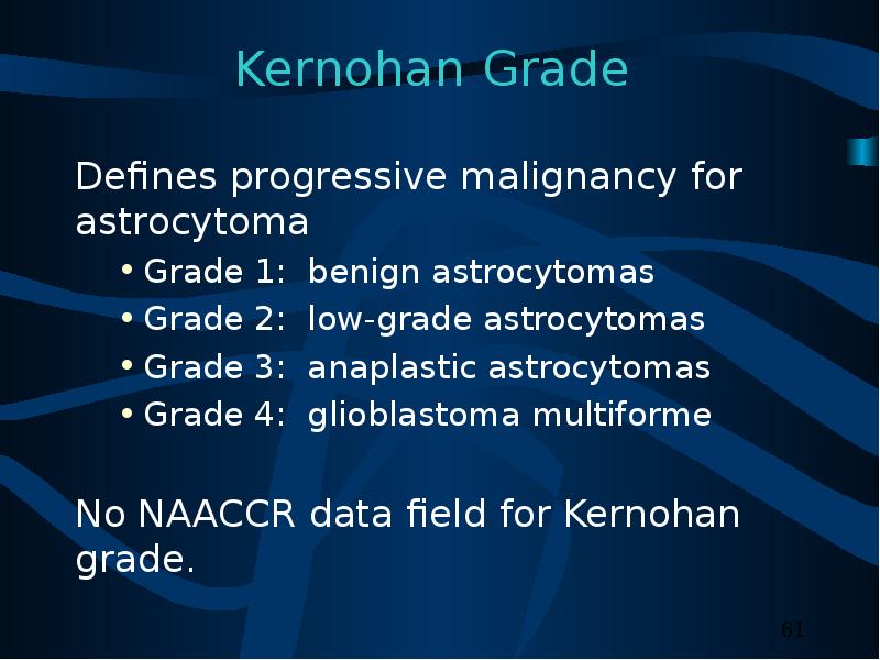
Содержание слайда: Kernohan Grade
Defines progressive malignancy for astrocytoma
Grade 1: benign astrocytomas
Grade 2: low-grade astrocytomas
Grade 3: anaplastic astrocytomas
Grade 4: glioblastoma multiforme
No NAACCR data field for Kernohan grade.
№62 слайд
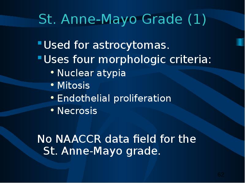
Содержание слайда: St. Anne-Mayo Grade (1)
Used for astrocytomas.
Uses four morphologic criteria:
Nuclear atypia
Mitosis
Endothelial proliferation
Necrosis
No NAACCR data field for the St. Anne-Mayo grade.
№63 слайд
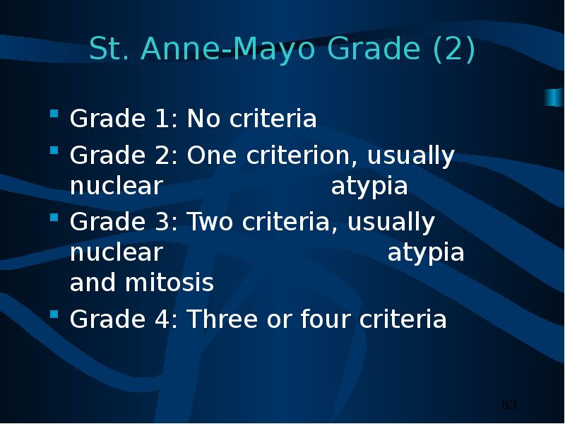
Содержание слайда: St. Anne-Mayo Grade (2)
Grade 1: No criteria
Grade 2: One criterion, usually nuclear atypia
Grade 3: Two criteria, usually nuclear atypia and mitosis
Grade 4: Three or four criteria
№64 слайд
Содержание слайда: Grade for CNS Tumors
Do not record WHO grade, Kernohan grade, or St. Anne/Mayo grade in the sixth digit histology code data field
№65 слайд

Содержание слайда: Part III
Laterality
Multiple Primaries
Malignant Transformation
Sequence Numbers
Date of Diagnosis
№66 слайд

Содержание слайда: Determining Multiple Primaries:
Laterality
Brain is not a paired organ.
Laterality collected on both non-malignant and malignant tumors.
Used to determine if multiple non-malignant CNS tumors are counted as multiple primary tumors.
Not used to determine if multiple malignant tumors of the same intracranial or CNS site are multiple primary tumors.
№67 слайд

Содержание слайда: Coding Laterality (1)
CNS sites to be coded with laterality:
Cerebral meninges, NOS (C70.0)
Cerebrum (C71.0)
Frontal lobe (C71.1)
Temporal lobe (C71.2)
Parietal lobe (C71.3)
Occipital lobe (C71.4).
№68 слайд
Содержание слайда: Coding Laterality (2)
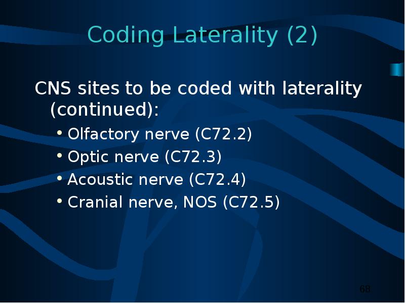
CNS sites to be coded with laterality (continued):
Olfactory nerve (C72.2)
Optic nerve (C72.3)
Acoustic nerve (C72.4)
Cranial nerve, NOS (C72.5)
№69 слайд

Содержание слайда: Determining Multiple Primaries:
Definitions
Non-malignant tumor
Tumor with ICD-O-3 behavior code
0 (benign) or 1 (borderline).
CNS
Includes intracranial and central nervous system topographic sites.
№70 слайд

Содержание слайда: Determining Multiple Primaries
Malignant (1)
NO CHANGES (at this time)
Site
Rule: Each category (first three characters) as delineated in ICD-O-3 is considered to be a separate site.
Multiple tumors are:
Same: C71.0 Cerebrum, C71.2 Temporal lobe
Different: C70.0 Cerebral Meninges, C71.0 Cerebrum
№71 слайд

Содержание слайда: Determining Multiple Primaries:
Malignant (2)
Histology
Rule: Differences in histologic type refer to differences in the FIRST THREE digits of the morphology code.
Multiple tumors in the same site are:
Same: Choroid plexus carcinoma (M9390), Ependymoma (M9391)
Different: Astrocytoma (M9400), Gemistocytic astrocytoma (M9411)
№72 слайд

Содержание слайда: Determining Multiple Primaries
Non-malignant (1)
NEW RULES
Site
Rule: Each sub-site (fourth-digit level) as delineated in ICD-O-3 is considered a separate site.
Same site if separate tumors with the same histology are in the same sub-site.
Different site if separate tumors have the same histology in different sub-site
C71.1 Frontal lobe, C71.4 Occipital lobe
C70.0 Cerebral Meninges, C70.1 Spinal meninges.
№73 слайд
Содержание слайда: Determining Multiple Primaries
Non-malignant (2)
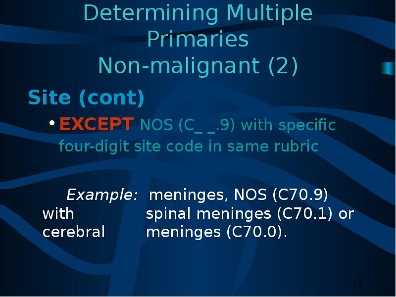
Site (cont)
EXCEPT NOS (C_ _.9) with specific four-digit site code in same rubric
Example: meninges, NOS (C70.9) with spinal meninges (C70.1) or cerebral meninges (C70.0).
№74 слайд

Содержание слайда: Determining Multiple Primaries
Non-malignant (3)
Site (cont)
Laterality: For non-malignant cases only
If multiple tumors of the same site and same histologic type are identified and both sides of a site listed as lateral are involved, tumors should be counted as separate primaries.
Different:
Right temporal lobe (C71.2) and left temporal lobe (C71.2)
№75 слайд

Содержание слайда: Determining Multiple Primaries:
Non-malignant (4)
№76 слайд

Содержание слайда: Determining Multiple Primaries:
Non-malignant (5)
Histology
If multiple tumors are in the same site, refer to Table 2, and use the following rules in priority order:
A-1: If the first three digits are the same but the codes are not found in Table 2, then the histology is considered to be the SAME.
A-2: If the first three digits are different but the codes are not found in Table 2, then the histology is considered to be DIFFERENT.
№77 слайд

Содержание слайда: Determining Multiple Primaries:
Non-malignant (6)
Histology (cont.)
B. If all histologies are listed in the same histologic group in Table 2, then the histology is considered to be the SAME. *
Example: Ependymomas: M9394, Myxopapillary ependymoma and M9444, Chordoid glioma have the same histology
*Note: If two histologies are in the same group in Table 2, code the first or more specific histology.
№78 слайд

Содержание слайда: Determining Multiple Primaries:
Non-malignant (7)
Histology (cont)
C: If the first three digits are the same as the first three digits for any histology in one of the groupings in Table 2 , then the histology is considered to be the SAME.*
Example: On table: Neuronal and neurol-glial neoplasm: M9505, ganglioglioma, Not on table: M9507, Pacinian tumor
* Note: If two histologies are in the same group in Table 2, code the first or more specific histology.
№79 слайд
Содержание слайда: Determining Multiple Primaries:
Non-malignant (8)
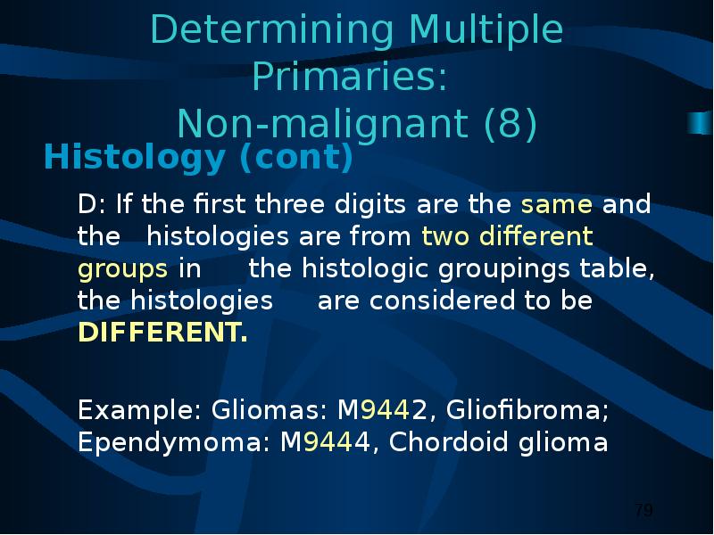
Histology (cont)
D: If the first three digits are the same and the histologies are from two different groups in the histologic groupings table, the histologies are considered to be DIFFERENT.
Example: Gliomas: M9442, Gliofibroma; Ependymoma: M9444, Chordoid glioma
№80 слайд
Содержание слайда: Determining Multiple Primaries:
Timing (1)
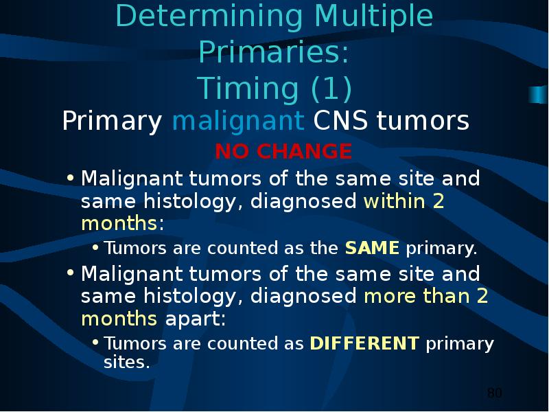
Primary malignant CNS tumors
NO CHANGE
Malignant tumors of the same site and same histology, diagnosed within 2 months:
Tumors are counted as the SAME primary.
Malignant tumors of the same site and same histology, diagnosed more than 2 months apart:
Tumors are counted as DIFFERENT primary sites.
№81 слайд
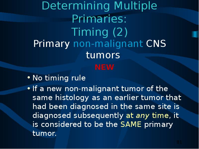
Содержание слайда: Determining Multiple Primaries:
Timing (2)
Primary non-malignant CNS tumors
NEW
No timing rule
If a new non-malignant tumor of the same histology as an earlier tumor that had been diagnosed in the same site is diagnosed subsequently at any time, it is considered to be the SAME primary tumor.
№82 слайд
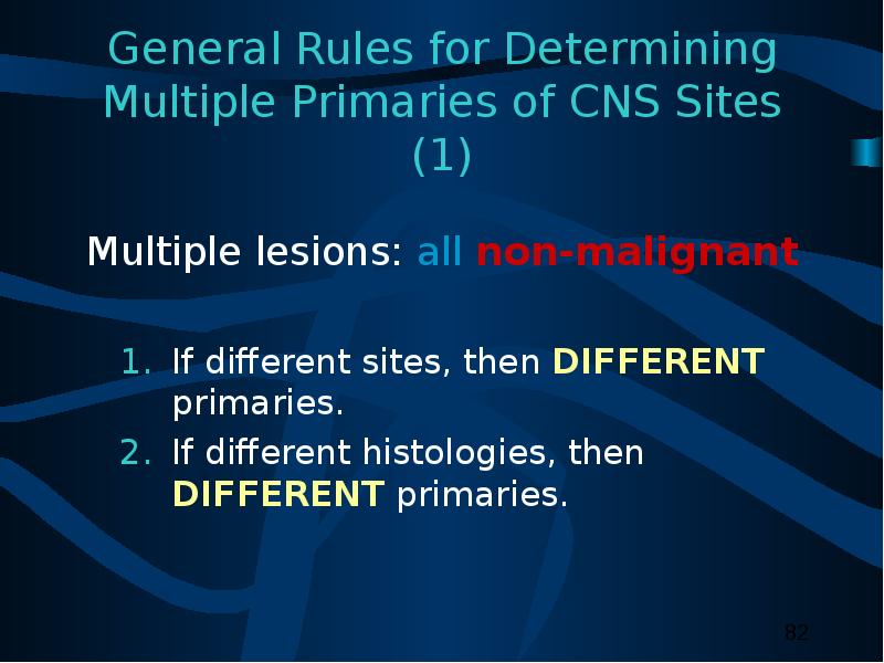
Содержание слайда: General Rules for Determining Multiple Primaries of CNS Sites (1)
Multiple lesions: all non-malignant
If different sites, then DIFFERENT primaries.
If different histologies, then DIFFERENT primaries.
№83 слайд

Содержание слайда: General Rules for Determining Multiple Primaries of CNS Sites (2)
Multiple lesions: all non-malignant (cont.)
If same site and same histology:
Laterality is same side, one side unknown or not applicable, then SAME primary.
Laterality is both sides, then DIFFERENT primaries.
№84 слайд
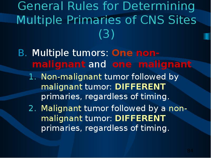
Содержание слайда: General Rules for Determining Multiple Primaries of CNS Sites (3)
Multiple tumors: One non-malignant and one malignant
Non-malignant tumor followed by malignant tumor: DIFFERENT primaries, regardless of timing.
Malignant tumor followed by a non-malignant tumor: DIFFERENT primaries, regardless of timing.
№85 слайд

Содержание слайда: Histologic Transformation (1)
Histologic transformation or progression to a higher grade:
Determined by pathological review.
Final diagnosis made by review of previous biopsies or excisions and comparison to newly biopsied or resected brain tumor
Non-malignant tumor transforms to malignant tumor.
Malignant tumors transforms to higher grade tumor.
№86 слайд
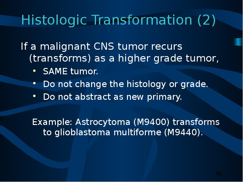
Содержание слайда: Histologic Transformation (2)
If a malignant CNS tumor recurs (transforms) as a higher grade tumor,
SAME tumor.
Do not change the histology or grade.
Do not abstract as new primary.
Example: Astrocytoma (M9400) transforms to glioblastoma multiforme (M9440).
№87 слайд

Содержание слайда: Histologic Transformation (3)
Transformation of a non-malignant tumor to a malignant tumor is rare.
Malignant transformations include:
Changes from WHO grade I to WHO grade II, III, or IV.
Changes from behavior code 0 or 1 to code 2 or 3.
Complete two abstracts:
One for the non-malignant tumor
One for the malignant tumor
№88 слайд
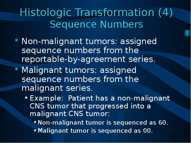
Содержание слайда: Histologic Transformation (4)
Sequence Numbers
Non-malignant tumors: assigned sequence numbers from the reportable-by-agreement series.
Malignant tumors: assigned sequence numbers from the malignant series.
Example: Patient has a non-malignant CNS tumor that progressed into a malignant CNS tumor:
Non-malignant tumor is sequenced as 60.
Malignant tumor is sequenced as 00.
№89 слайд

Содержание слайда: Histologic Transformation (5)
Date of Diagnosis
Non-malignant tumors: First date that a medical practitioner diagnosed the non-malignant tumor either clinically or histologically.
Malignant tumors: First date that a medical practitioner diagnosed the malignant transformation either clinically or histologically.
№90 слайд

Содержание слайда: Coding Sequence Numbers (1)
Indicates the sequence of all reportable neoplasms over the lifetime of the person.
Codes 00 – 35: Malignant and in situ reportable neoplasms.
Codes 60 – 87: Reportable-by-agreement including non-malignant tumors diagnosed after January1, 2004.
№91 слайд

Содержание слайда: Coding Sequence Numbers (2)
Reportable-by-agreement neoplasms are defined by each facility and/or central cancer registry:
Non-malignant CNS tumors are assigned reportable-by-agreement sequence numbers even when they are reportable.
Codes 60 – 87
№92 слайд
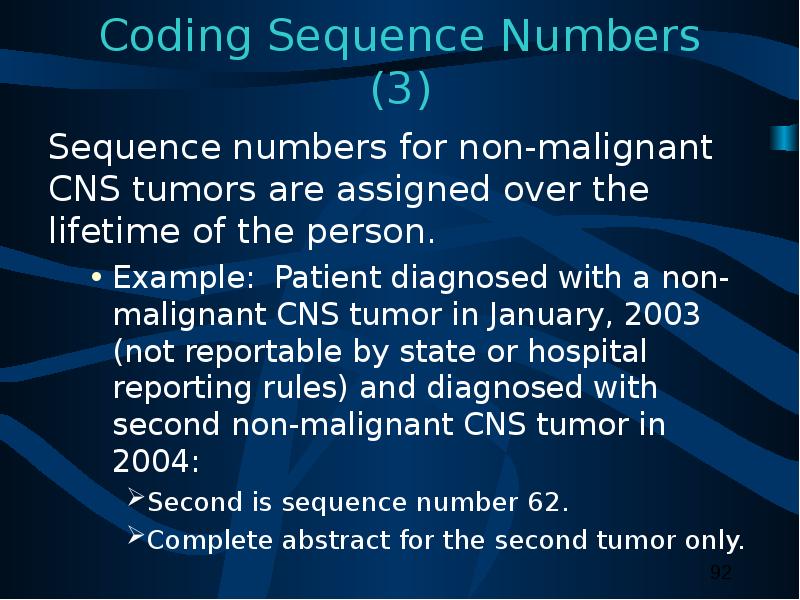
Содержание слайда: Coding Sequence Numbers (3)
Sequence numbers for non-malignant CNS tumors are assigned over the lifetime of the person.
Example: Patient diagnosed with a non-malignant CNS tumor in January, 2003 (not reportable by state or hospital reporting rules) and diagnosed with second non-malignant CNS tumor in 2004:
Second is sequence number 62.
Complete abstract for the second tumor only.
№93 слайд

Содержание слайда: Assigning Diagnosis Date
Rules for assigning diagnosis date are the same for malignant and non-malignant tumors.
Review source records carefully to determine initial diagnosis date, regardless of whether it is a clinical or histological diagnosis.
№94 слайд

Содержание слайда: Part IV
Staging
Risk Factors
Genetic Syndromes
Diagnostic Tools
Treatment
Edits
Data Analysis
№95 слайд

Содержание слайда: Collaborative Stage (CS)
A computer algorithm uses the collaborative stage (CS) data fields to calculate site-specific American Joint Committee on Cancer (AJCC) TNM stage, SEER Summary Stage 1977, and SEER Summary Stage 2000.
№96 слайд
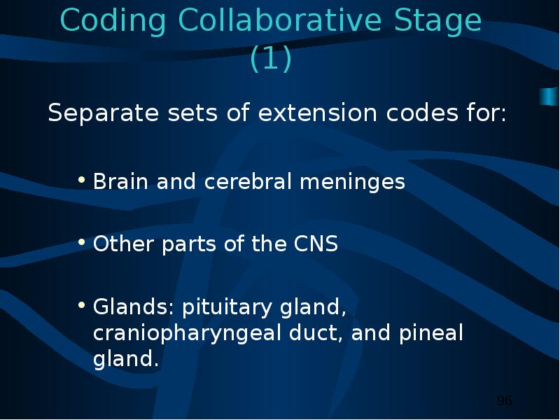
Содержание слайда: Coding Collaborative Stage (1)
Separate sets of extension codes for:
Brain and cerebral meninges
Other parts of the CNS
Glands: pituitary gland, craniopharyngeal duct, and pineal gland.
№97 слайд

Содержание слайда: Coding Collaborative Stage (2)
Site-specific codes for lymph nodes
Same for the Brain, cerebral meninges and other CNS.
Code 88: Not applicable.
For pituitary gland, craniopharyngeal duct, and pineal gland
Code 99: Not applicable.
Metastasis at Diagnosis
Same for the pituitary gland, craniopharyngeal duct, and pineal gland and other CNS.
Different for brain and cerebral meninges.
№98 слайд
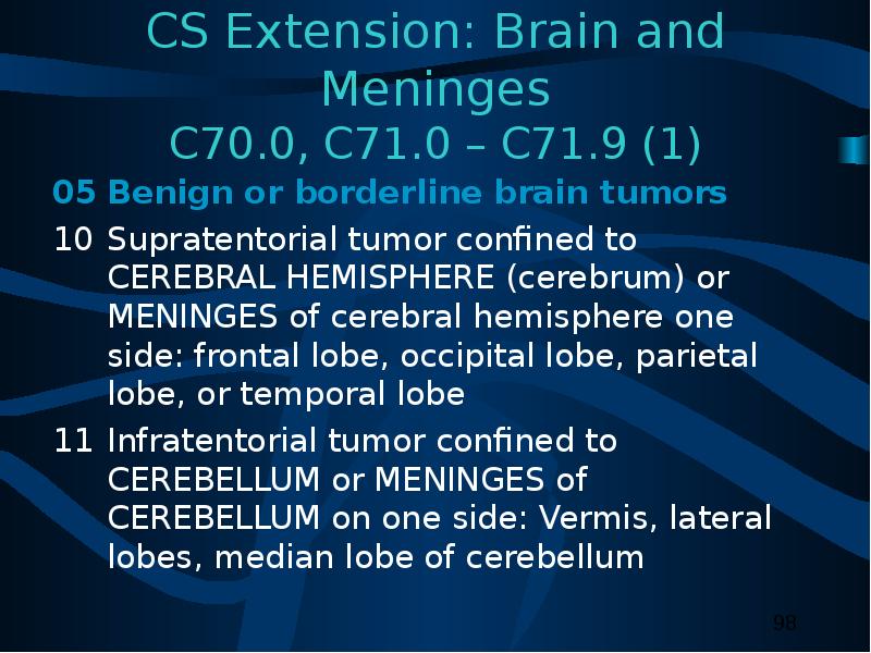
Содержание слайда: CS Extension: Brain and Meninges
C70.0, C71.0 – C71.9 (1)
05 Benign or borderline brain tumors
10 Supratentorial tumor confined to CEREBRAL HEMISPHERE (cerebrum) or MENINGES of cerebral hemisphere one side: frontal lobe, occipital lobe, parietal lobe, or temporal lobe
11 Infratentorial tumor confined to CEREBELLUM or MENINGES of CEREBELLUM on one side: Vermis, lateral lobes, median lobe of cerebellum
№99 слайд
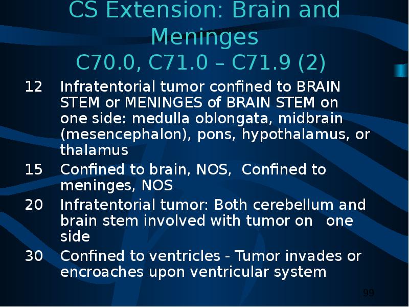
Содержание слайда: CS Extension: Brain and Meninges
C70.0, C71.0 – C71.9 (2)
12 Infratentorial tumor confined to BRAIN STEM or MENINGES of BRAIN STEM on one side: medulla oblongata, midbrain (mesencephalon), pons, hypothalamus, or thalamus
15 Confined to brain, NOS, Confined to meninges, NOS
20 Infratentorial tumor: Both cerebellum and brain stem involved with tumor on one side
30 Confined to ventricles - Tumor invades or encroaches upon ventricular system
№100 слайд
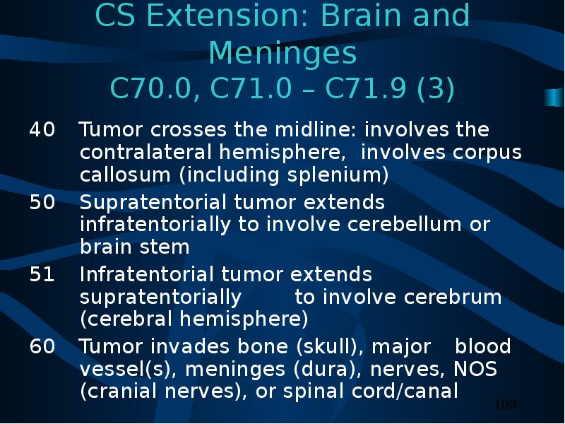
Содержание слайда: CS Extension: Brain and Meninges
C70.0, C71.0 – C71.9 (3)
40 Tumor crosses the midline: involves the contralateral hemisphere, involves corpus callosum (including splenium)
50 Supratentorial tumor extends infratentorially to involve cerebellum or brain stem
51 Infratentorial tumor extends supratentorially to involve cerebrum (cerebral hemisphere)
60 Tumor invades bone (skull), major blood vessel(s), meninges (dura), nerves, NOS (cranial nerves), or spinal cord/canal
№101 слайд

Содержание слайда: CS Extension: Brain and Meninges
C70.0, C71.0 – C71.9 (4)
70 Circulating cells in cerebral spinal fluid; nasal cavity; nasopharynx; posterior pharynx; or outside CNS
80 Further contiguous extension
95 No evidence of primary tumor
99 Unknown extension; Primary tumor cannot be accessed; Not documented in patient record
№102 слайд
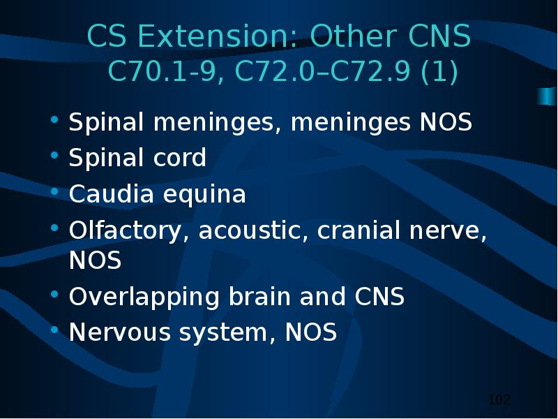
Содержание слайда: CS Extension: Other CNS
C70.1-9, C72.0–C72.9 (1)
Spinal meninges, meninges NOS
Spinal cord
Caudia equina
Olfactory, acoustic, cranial nerve, NOS
Overlapping brain and CNS
Nervous system, NOS
№103 слайд

Содержание слайда: CS Extension: Other CNS
C70.1-9, C72.0–C72.9 (2)
05 Benign or borderline tumors
10 Tumor confined to tissue or site of origin
30 Localized, NOS
40 Meningeal tumor infiltrates nerve; nerve tumor infiltrates meninges (dura)
50 Adjacent connective/soft tissue; adjacent muscle
60 Brain, for cranial nerve tumors; major blood vessel(s); sphenoid and frontal sinuses (skull)
№104 слайд

Содержание слайда: CS Extension: Other CNS
C70.1-9, C72.0–C72.9 (3)
70 Brain except for cranial nerve tumors; bone, other than skull; eye
80 Further contiguous extension
95 No evidence of primary tumor
99 Unknown extension; primary tumor cannot be assessed; not documented in patient record
№105 слайд

Содержание слайда: CS Extension: Other Endocrine
C75.1, C75.2, C75.3
00 In situ; non-invasive; intraepithelial
05 Benign or borderline tumors
10 Invasive carcinoma confined to gland of origin
30 Localized, NOS
40 Adjacent connective tissue
60 Pituitary and craniopharyngeal duct: Cavernous sinus; infundibulum; pons; sphenoid body and siunses
Pineal: Infratentorial and central brain
80 Further contiguous extension
95 No evidence of primary tumor
99 Unknown extension
№106 слайд

Содержание слайда: CS Lymph Nodes
Describes tumor involvement of regional lymph nodes.
Code for CS Lymph Nodes is 88 (not applicable) for meninges, brain, spinal cord, cranial nerves, and other parts of the CNS.
Code for CS Lymph Nodes is 99 (unknown, not stated) for pituitary gland, craniopharyngeal duct, and pineal gland.
№107 слайд

Содержание слайда: CS Metastasis at Diagnosis
Brain and Meninges
C70.0, C71.0-9
00 No; None
10 Distant metastases
85 “Drop” metastases
99 Unknown; distant metastasis cannot be assessed; not documented in patient record
№108 слайд

Содержание слайда: CS Metastasis at Diagnosis
Other CNS and Other Endocrine
C70.1-9, C72.0—9, C75.1, C75,2, C75.3
00 No; None
10 Distant lymph node(s)
40 Distant metastasis except lymph nodes (code 10)
Distant metastasis, NOS
Carcinomatosis
50 (40) + (10)
99 Unknown; distant metastasis cannot be assessed; not documented in patient record
№109 слайд
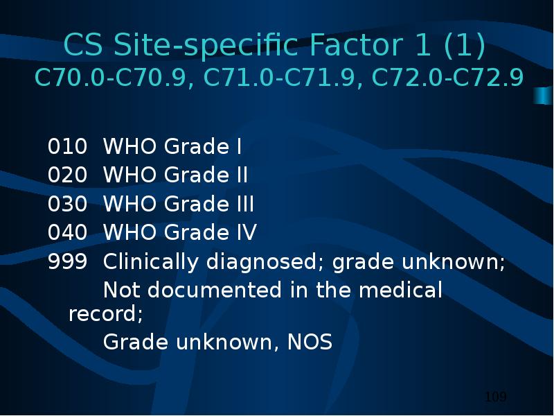
Содержание слайда: CS Site-specific Factor 1 (1)
C70.0-C70.9, C71.0-C71.9, C72.0-C72.9
010 WHO Grade I
020 WHO Grade II
030 WHO Grade III
040 WHO Grade IV
999 Clinically diagnosed; grade unknown;
Not documented in the medical record;
Grade unknown, NOS
№110 слайд

Содержание слайда: CS Site-specific Factor 1 (2)
C70.0-C70.9, C71.0-C71.9, C72.0-C72.9
C75.1- C75.3
Code the WHO grade for CNS tumors in CS Site-specific factor 1.
Do not code WHO grade in the sixth digit histology data field.
№111 слайд
Содержание слайда: Possible Risk Factors
Genetic predispositions for the development of brain tumors have been identified.
Population-based studies suggest that no more than 4% are attributed to heredity.
Several environmental factors that may be associated with CNS tumors.
№112 слайд

Содержание слайда: Possible Risk Factors
Epstein-Barr virus in the DNA of primary lymphoma suggests a viral etiology for CNS tumors.
Reference: “Surveillance of Primary Intracranial and Central Nervous System Tumors: Recommendations from the Brain Tumor Working Group.”
№113 слайд

Содержание слайда: Genetic Syndromes
Genetic syndromes associated with multiple CNS tumors are:
Neurofibromatosis I (von Recklinghausen’s disease)
Neurofibromatosis II (bilateral acoustic neurofibromatosis)
Von Hippel-Lindau disease
Tuberous sclerosis (Bourneville-Pringle syndrome)
Gorlin syndrome (Nevoid Basal Cell Carcinoma syndrome
Hermans-Grosfeld-Spaas-Valk disease
Li-Fraumeni syndrome
Familial retinoblastoma
Turcot syndrome (Adenomatous Polyposis syndrome)
Cowden disease
№114 слайд

Содержание слайда: Diagnostic Tools – Physical Exam
Neurological examination
Eye movements
Vision
Hearing
Reflexes
Balance and coordination
Sense of smell and touch
Abstract thinking
Memory
№115 слайд

Содержание слайда: Diagnostic Tools: Radiology
Computerized tomography (CT) scan
Magnetic resonance imaging (MRI)
Positron emission tomography (PET)
Single photon emission computed tomography (SPECT)
Magnetoencephalography (MEG)
Angiography
№116 слайд

Содержание слайда: Diagnostic Tools: Laboratory tests
Audiometry
Electroencephalogram (EEG)
Endocrine evaluation
Evoked potentials
Lumbar puncture
Myelogram
Perimetry
№117 слайд
Содержание слайда: Diagnostic Tools
Needle biopsy
Needle inserted through a burr hole and tissue extracted for tissue diagnosis.
Stereotactic biopsy
Computer used to guided needle biopsy to extract tissue.
№118 слайд
Содержание слайда: College of American Pathologist
(CAP) Protocols
Site-specific checklists
Required to be completed in the health record in hospitals with COC-approved cancer programs for cases diagnosed January 1, 2004 and later.
№119 слайд
Содержание слайда: Brain and Spinal Cord
CAP Protocols (1)
Macroscopic
Specimen type
Specimen size
Tumor site
Tumor size
№120 слайд

Содержание слайда: Brain and Spinal Cord
CAP Protocols
Microscopic
Histologic type
Histologic grade
Margins
Additional studies*
Additional pathologic findings*
Comments*
*Not required for COC approval.
№121 слайд
Содержание слайда: Treatment (1)
Watchful waiting
Surgery
Radiation
Chemotherapy
Hormonal therapy
Immunotherapy
Hematologic Transplant
and Endocrine procedures
№122 слайд
Содержание слайда: Treatment (2)
Inoperable or inaccessible tumors may be treated with primary radiation and other systemic therapy:
Chemotherapy, immunotherapy, and hormone therapy.
Shunt insertion to reduce intracranial swelling is not coded as surgical treatment.
№123 слайд
Содержание слайда: Surgical Procedure of Primary Site
Brain: Site-specific surgery codes
Meninges
Brain
Spinal cord, cranial nerves, other CNS.
№124 слайд

Содержание слайда: Surgical Procedure of Primary Site
C70-0-C70.9, C71.0-C71.9, C72.0-C72.9 (1)
Code 10: Tumor destruction, NOS
Laser surgery
Laser surgery with photodynamic therapy
Ultrasonic aspirator.
No specimen sent to pathology from surgical procedure.
№125 слайд

Содержание слайда: Surgical Procedure of Primary Site
C70-0-C70.9, C71.0-C71.9, C72.0-C72.9 (2)
20:Local Excision (biopsy) of tumor, lesion, or mass
Specimen sent to pathology from surgical event.
40: Partial resection
55: Gross total resection
90: Surgery, NOS
№126 слайд

Содержание слайда: Surgical Procedure of Primary Site
C75.1, C75.2, C75.3 (1)
Code 10: Local tumor destruction, NOS
Code 11: Photodynamic therapy
Code 12: Electrocautery; fulguration
Code 13: Cryosurgery
Code 14: Laser
No specimen is sent to pathology from surgical events 10-14.
№127 слайд

Содержание слайда: Surgical Procedure of Primary Site
C75.1, C75.2, C75.3 (2)
Code 20: Local tumor excision, NOS
Code 26: Polypectomy
Code 27: Excisional biopsy
Any combination of 20 or 26-27 WITH
21: Photodynamic therapy (PDT)
22: Electrocautery
23: Cyrosurgery
24: Laser ablation
№128 слайд

Содержание слайда: Surgical Procedure of Primary Site
C75.1, C75.2, C75.3 (3)
Code 25: Laser excision
Specimen sent to pathology from surgical event 20-27.
Code 30: Simple or partial surgical removal of primary site.
№129 слайд

Содержание слайда: Surgical Procedure of Primary Site
C75.1, C75.2, C75.3 (4)
Code 40: Total surgical removal of primary site; enucleation
Code 50: Surgery stated to be “debulking”
Code 60: Radical surgery
Partial or total removal of the primary site WITH resection in continuity (partial or total removal) with other organs
Code 90: Surgery, NOS
№130 слайд
Содержание слайда: Surgical Margins of the Primary Site
Code final status of surgical margins
COC-required data item.
Serves as quality control measure for pathology reports.
May be prognostic factor in recurrence.
№131 слайд

Содержание слайда: Scope of Regional Lymph Node Surgery
Identifies removal, biopsy, or aspiration of regional lymph node(s):
NPCR-, COC-, and SEER-required data item.
Code 9: Meninges, brain, and spinal cord; cranial nerves; and other parts of the CNS.
Code as appropriate: Pituitary gland, craniopharyngeal duct, and pineal gland.
№132 слайд
Содержание слайда: Radiation Therapy (1)
Radiation codes indicate type of radiation therapy performed as part of the first course of treatment.
Records modality of radiation therapy used to deliver significant regional dose to the primary volume of interest.
COC-required data item.
SEER collects these data from COC-approved facilities
NPCR: Supplementary or recommended.
№133 слайд

Содержание слайда: Radiation Therapy (2)
Beam radiation
Codes 20 – 29:
Conventional radiation therapy: from an external beam directed at the tumor.
Energy is orthovoltage, cobalt, photon, and/or electron.
Code 30: Boron neutron capture therapy (BNCT)
Code 31: Intensity-modulated radiation therapy (IMRT)
№134 слайд

Содержание слайда: Radiation Therapy (3)
Beam radiation
Code 32: Conformal radiation
Code 40: Particle or proton beam
Code 41: Stereotactic radiosurgery, NOS
Code 42: Linac radiosurgery
Code 43: Gamma knife
№135 слайд

Содержание слайда: Radiation Therapy (3)
Tumors typically treated with stereotactic radiosurgery include:
№136 слайд

Содержание слайда: Radiation Therapy (4)
Radioactive implants
Code 50: Brachytherapy, radiation implants, radiation seeding, radioactive implants, interstitial implants, intracavitary radiation NOS
Code 51: Intracavitary radiation with low dose rate applicators (Cesium- 137, Fletcher applicator)
№137 слайд
Содержание слайда: Radiation Therapy (5)
Radioactive implants (continued)
Code 52: Intracavitary radiation with high dose rate applicator
Code 53: Interstitial radiation with low dose rate sources
Code 54: Interstitial radiation with high dose rate sources
Code 55: Low dose rate interstitial or intracavitary radium
№138 слайд

Содержание слайда: Chemotherapy (1)
Record type of chemotherapy administered as first course of treatment:
Code 01: Chemotherapy, NOS
Code 02: Single-agent chemotherapy
Code 03: Multi-agent chemotherapy
№139 слайд
Содержание слайда: Chemotherapy (2)
Blood-brain barrier
Protects the brain from foreign substances, including chemotherapy.
May be disrupted by receptor-mediated permeabilizers.
Intrathecal chemotherapy
Drugs directly injected into the cerebrospinal fluid by spinal injection or Ommaya reservoir.
№140 слайд

Содержание слайда: Chemotherapy (3)
Interstitial chemotherapy
Administered directly to involved tissues.
Polymer wafers soaked in a chemotherapeutic agent are inserted in the tumor bed after tumor resection.
№141 слайд
Содержание слайда: Hormone Therapy
Record systemic hormonal agents administered as first course of treatment.
Tamoxifen and RU-486 (Mifepristone) may be used to treat meningioma.
Steroids given to treat swelling caused by CNS tumors are not coded as hormone therapy.
№142 слайд

Содержание слайда: Immunotherapy (1)
Record whether immunotherapeutic agents were administered as first course of treatment:
Angiogenesis inhibitors block the development of new blood vessels and starve the tumor.
Interleukins are growth factors that manipulate the tumor’s ability to grow.
№143 слайд

Содержание слайда: Immunotherapy (2)
Gene therapy replaces or repairs the gene responsible for tumor growth.
Vaccine therapy allows the immune system to detect the tumor antigens and attack the tumor cells.
№144 слайд

Содержание слайда: Hematologic Transplant and Endocrine Procedures
Identify systemic therapeutic procedures administered as first course of treatment:
Code 10: Bone marrow transplant, NOS
Code 11: Autologous bone marrow transplant
Code 12: Allogeneic bone marrow transplant
Code 20: Stem cell harvest
Code 30: Endocrine surgery and/or endocrine radiation therapy
Code 40: Combination of endocrine surgery and/or radiation with transplant procedure
№145 слайд
Содержание слайда: Technical Issues
Edit Checks
NAACCR Edits Committee is developing and modifying data edits to accommodate data collection of non-malignant CNS tumors.
№146 слайд
Содержание слайда: Technical Issues
Data Analysis Recommendations
Report and analyze data for non-malignant CNS tumors separately from malignant tumors.
Footnote that pilocytic astrocytomas are included in the analysis for malignant brain tumors for continuity of trends.
Review the standard site and histology groupings for tabulating estimates of these tumors to allow comparability of information across registries.
№147 слайд
Содержание слайда: References
Manuals, Articles, Reports
A Primer of Brain Tumors, 1998; American Brain Tumor Association, Des Plaines, IL; 800-886-2282 (can link to the manual through their website: www.abta.org)
Gershman S, Surawicz T, McLaughlin V, Rousseau V. Completeness of Reporting of Brain and Other Central Nervous System Neoplasms. Journal of Registry Management, Winter 2001, Volume 28, Number 4.
№148 слайд

Содержание слайда: References
Manuals, Articles, Reports (continued)
Fritz A, Percy C, Jack V, Shanmugaratnam K, Sobin V, Parkin D M , Whelan S. International Classification of Diseases for Oncology, 3rd ed. Geneva: World Health Organization, 2000
Report: Surveillance of Primary Intracranial and Central Nervous System Tumors: Recommendations from the Brain Tumor Working Group, National Coordinating Council for Cancer Surveillance, September 1998
№149 слайд

Содержание слайда: References
Websites
American Brain Tumor Association www.abta.org
American College of Surgeons, Commission on Cancer Information, Facility Oncology Data Standards (FORDS) www.facs.org/dept/cancer/index.html
American Joint Committee on Cancer, Collaborative Stage Documentation www.edits.cx/cs/
№150 слайд

Содержание слайда: References
Websites (continued)
Brain and Neurosurgery Information Center www.brain-surgery.com/index.html
Brain and Spinal Cord Tumors: Hope through Research www.ninds.nih.gov/health_and_medical/pubs/brain_tumor_hope_through_research.htm
Brain Tumor Guide http://virtualtrials.com/faq/toc.cfm
Central Brain Tumor Registry of the United States www.cbtrus.org/page2t.htm
№151 слайд
Содержание слайда: References
Websites (continued)
College of American Pathologists (CAP), Protocol – Brain ftp://ftp.cap.org/cancerprotocols/Brain03_p.doc
Illustrated Glossary of Radiology: Anatomy, Examinations and Procedures; Department of Radiology and Radiological Services, The Uniformed Services University of the Health Sciences
http://rad.usuhs.mil/glossary.html
№152 слайд

Содержание слайда: References
Websites (continued)
International RadioSurgery Association www.isra.org/index.html
National Brain Tumor Radiosurgery Association www.braintumors.com/radiosurgery/radiosrugery.info#TWO
NCI Brain Tumor Home Page www.nci.nih.gov/cancer_information/cancer_type/brain_tumor/
№153 слайд

Содержание слайда: References
Websites (continued)
PDQ Cancer Information Summaries: Adult Treatment www.cancer.gov/cancerinfo/pdq/adulttreatment
PDQ Cancer Information Summaries: Pediatric Treatment www.cancer.gov/cancerinfo/pdq/pediatrictreatment
The Brain Tumor Foundation www.braintumorfoundation.org/neurosurgery/ss3_3.htm
№154 слайд
Содержание слайда: Acknowledgments (1)
Prepared by
Shannon Vann, CTR
for the
North American Association of Central Cancer Registries (NAACCR)
This training presentation was supported by contract #200-2001-00044 from CDC. The content of this training presentation does not necessarily reflect the views or policies of the Department of Health and Human Services, nor does mention of trade names, commercial products, or organizations imply endorsement by the U.S. Government.
№155 слайд
Содержание слайда: Acknowledgments (2)
Sponsors
Centers for Disease Control and Prevention
National Program for Cancer Registries
National Cancer Institute
Surveillance, Epidemiology and End Results Program
North American Association of Central Cancer Registries
American Joint Committee on Cancer
Collaborative Stage Task Force
№156 слайд
Содержание слайда: Acknowledgments (3)
CDC National Program of Cancer Registries Planning Committee
Kimberly Cantrell
Gayle G. Clutter
Faye Floyd
Michael Lanzilotta
Frances Michaud
№157 слайд

Содержание слайда: Acknowledgments (4)
Materials Review Committee
Trista Aarnes-Leong St. Vincent Medical Center, NAACCR Registry Operations Subcommittee,
Susan Bolick-Aldrich South Carolina Central Cancer Registry, NAACCR Registry Operations Subcommittee, Chair, Co-chair, Registry Operations Committee
Gayle Clutter CDC National Program of Cancer Registries, Registry Operations Subcommittee, National Coordination Council on Cancer Surveillance Brain Tumor Working Group, Chair
Faye Floyd CDC National Program of Cancer Registries
April Fritz NCI Surveillance, Epidemiology and End Results Program, Registry Operations Subcommittee
Elaine Hamlyn Canadian Cancer Registry, Registry Operations Subcommittee,
Holly Howe North American Association of Central Cancer Registries, Executive Director
Betsy Kohler New Jersey State Cancer Registry, NAACCR Education Committee
Carol Kruchko Central Brain Tumor Registry of the United States, Registry Operations Subcommittee, National Coordination Council on Cancer Surveillance Brain Tumor Working Group
Donna Morrel Cancer Surveillance Program of Los Angeles. Registry Operations Subcommittee
Linda Mulvihill North Carolina Central Cancer Registry, Registry Operations Subcommittee
Wendy Scharber Minnesota Cancer Surveillance Program
James Smirniotopoulos Professor of Radiology, Uniformed Services University, Registry Operations Subcommittee
Katheryne Vance California Cancer Registry, Registry Operations Subcommittee
Valerie Vesich American College of Surgeons, Commission on Cancer, Registry Operations Subcommittee
Скачать все slide презентации Data Collection of Primary Central Nervous System (CNS) Tumors одним архивом: